
 By Nana O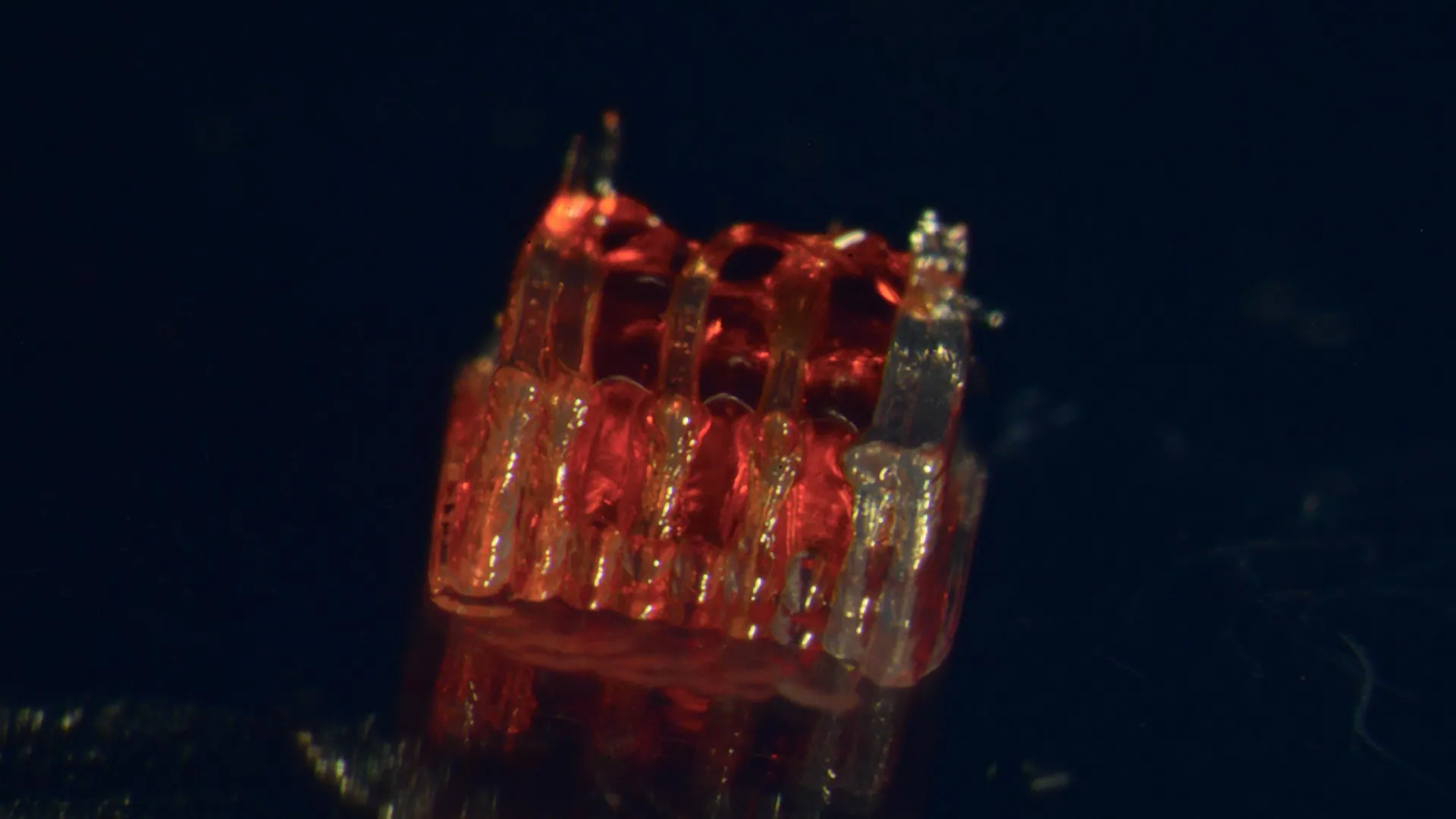
A pioneering research endeavor at the University of Minnesota Twin Cities has achieved a significant milestone in the quest for spinal cord injury recovery, marking the first instance of successfully integrating 3D printing technology with stem cell biology and the cultivation of lab-grown tissues. This groundbreaking process offers a novel approach to addressing the devastating consequences of spinal cord damage, a condition that affects hundreds of thousands of individuals annually. The findings of this transformative study were recently disseminated in the esteemed, peer-reviewed scientific journal Advanced Healthcare Materials.
The pervasive challenge of spinal cord injuries (SCIs) in the United States is underscored by sobering statistics. According to the National Spinal Cord Injury Statistical Center, more than 300,000 individuals are living with the profound and often irreversible effects of SCIs. Despite decades of intensive research, a complete reversal of the damage and subsequent paralysis remains an elusive goal. A primary impediment to recovery lies in the inherent vulnerability of nerve cells, which are prone to death following trauma, and the subsequent failure of nerve fibers to regenerate across the injured epicenter. This new research directly confronts these critical biological hurdles, presenting a potential pathway toward restoring lost function.
The innovative methodology developed by the University of Minnesota team centers on the creation of a sophisticated, three-dimensional printed framework, termed an organoid scaffold. This scaffold is meticulously designed with microscopic channels, serving as a blueprint for the development of lab-grown organoids. These intricate channels are then strategically populated with regionally specific spinal neural progenitor cells (sNPCs). These sNPCs are a specialized type of cell derived from human adult stem cells, possessing the remarkable capacity to proliferate and differentiate into various specific types of mature nerve cells.
Guebum Han, a former postdoctoral researcher in mechanical engineering at the University of Minnesota and the lead author of the published paper, who now contributes his expertise to Intel Corporation, elaborated on the precision of their approach. "We utilize the 3D printed channels within the scaffold to meticulously guide the growth of the stem cells," Dr. Han explained. "This directed growth is crucial for ensuring that the newly formed nerve fibers extend in the intended, functional manner." He further emphasized the functional outcome: "This method effectively constructs a sophisticated relay system. When this engineered construct is implanted into the spinal cord, it serves to bypass the damaged area, re-establishing vital communication pathways."
To rigorously test the efficacy of their novel approach, the researchers embarked on a series of preclinical trials. In their published study, these sophisticated scaffolds, teeming with sNPCs, were transplanted into rats that had sustained completely severed spinal cords. The results were remarkably promising. The implanted cells demonstrated a robust ability to differentiate into functional neurons. Crucially, these newly formed nerve fibers successfully extended their axons in both directions – rostrally (towards the head) and caudally (towards the tail) – thereby forming new and functional connections with the host’s pre-existing neural circuitry. This integration signifies a critical step in bridging the gap created by the injury.
The study meticulously documented the integration process over time. The new nerve cells not only survived but also seamlessly integrated into the host spinal cord tissue. This biological integration facilitated a significant and measurable functional recovery in the experimental subjects. The rats exhibited improvements in motor function and coordination, demonstrating the potential of this bioengineered intervention to restore lost capabilities.
Ann Parr, a professor of neurosurgery at the University of Minnesota and a key investigator in the research, articulated the broader significance of these findings within the field of regenerative medicine. "Regenerative medicine has undeniably ushered in a new era of exploration and innovation in spinal cord injury research," Professor Parr stated. "Our laboratory is profoundly enthusiastic about exploring the future potential of these ‘mini spinal cords’ and their prospects for clinical translation to human patients."
While acknowledging that the research is still in its nascent stages, the implications are far-reaching and offer a beacon of hope for individuals living with the debilitating effects of spinal cord injuries. The research team’s immediate objective is to scale up the production of these bioengineered scaffolds and to continue refining the synergistic combination of these advanced technologies. The ultimate aim is to pave the way for future clinical applications that could revolutionize SCI treatment.
The collaborative nature of this groundbreaking research is evident in the diverse expertise brought together by the University of Minnesota. Beyond Dr. Han and Professor Parr, the core research team included Hyunjun Kim and Michael McAlpine from the University of Minnesota Department of Mechanical Engineering, bringing their expertise in advanced manufacturing and biomechanics. Nicolas S. Lavoie, Nandadevi Patil, and Olivia G. Korenfeld from the University of Minnesota Department of Neurosurgery contributed their deep understanding of neurological pathways and surgical considerations. Manuel Esguerra from the University of Minnesota Department of Neuroscience provided critical insights into neural development and regeneration. Furthermore, Daeha Joung from the Department of Physics at Virginia Commonwealth University played a vital role in the physical characterization and engineering of the scaffold.
The significant financial and institutional backing for this complex research underscores its recognized importance. This work received crucial funding from the National Institutes of Health, a leading federal agency dedicated to advancing biomedical research. Additional support was provided by the State of Minnesota Spinal Cord Injury and Traumatic Brain Injury Research Grant Program, highlighting state-level commitment to addressing these critical health issues. The Spinal Cord Society also contributed funding, demonstrating broad-based support for innovative SCI research.
The full scientific exposition of this research, titled "3D-Printed Scaffolds Promote Enhanced Spinal Organoid Formation for Use in Spinal Cord Injury," is accessible on the Advanced Healthcare Materials website, providing a comprehensive resource for the scientific community.
Background and Context: The Enduring Challenge of Spinal Cord Injury
Spinal cord injuries are among the most devastating and life-altering medical conditions, resulting from damage to the intricate network of nerves that transmit signals between the brain and the rest of the body. The consequences can range from temporary loss of sensation and motor function to permanent paralysis, profoundly impacting an individual’s independence, quality of life, and overall well-being. The economic burden associated with SCI care is immense, encompassing lifelong medical expenses, rehabilitation services, assistive devices, and lost productivity.
Historically, the medical community has faced significant challenges in treating SCIs. The central nervous system, unlike other tissues in the body, has a limited capacity for self-repair. Once nerve cells are destroyed or nerve fibers are severed, they rarely regenerate spontaneously. This inherent biological limitation has made developing effective therapeutic strategies an uphill battle. Early research focused on managing symptoms, preventing secondary complications like infections and pressure sores, and maximizing remaining function through physical therapy.
In recent decades, the advent of regenerative medicine has ignited a new wave of optimism. This interdisciplinary field aims to restore the structure and function of damaged tissues and organs through various approaches, including cell-based therapies, tissue engineering, and the use of biomaterials. Stem cell research, in particular, has offered immense promise due to the potential of stem cells to differentiate into specialized cell types, including neurons. However, effectively delivering and guiding these cells to form functional neural circuits within the complex environment of the injured spinal cord has remained a formidable obstacle.
Timeline and Evolution of the Research
While the precise chronological development of this specific project is not detailed in the initial report, the University of Minnesota’s involvement in spinal cord injury research, particularly in the realms of neurosurgery, neuroscience, and bioengineering, has been ongoing. This current study likely represents a culmination of years of foundational research in stem cell biology, 3D printing technologies for biomedical applications, and preclinical animal models. The publication in Advanced Healthcare Materials suggests a rigorous peer-review process, indicating that the research has progressed through multiple stages of experimentation, data analysis, and validation. The evolution of this work can be seen as a progression from understanding the basic biological mechanisms of neural repair to developing sophisticated engineering solutions that mimic natural biological processes.
Supporting Data and Scientific Principles
The success of this research hinges on several key scientific principles:
- Stem Cell Differentiation: Human adult stem cells, specifically sNPCs, possess multipotency, meaning they can develop into a variety of specialized cell types, including the neurons essential for nerve signal transmission. This ability is fundamental to replacing damaged nerve cells.
- 3D Printing for Precision Engineering: 3D printing allows for the creation of highly intricate and customizable structures. In this context, the microscopic channels within the scaffold act as precise conduits, guiding the directional growth of neuronal axons, a critical factor for re-establishing functional connections. This overcomes the natural tendency of axons to grow in a disorganized manner after injury.
- Biocompatibility and Integration: For any implanted biomaterial or cell therapy to be successful, it must be biocompatible, meaning it does not elicit a detrimental immune response from the host. Furthermore, the engineered tissue must be capable of integrating with the host’s existing biological systems. The study’s finding of seamless integration in rats indicates successful biocompatibility and functional integration.
- Scaffold as a Bio-Mimetic Environment: The organoid scaffold is designed to mimic the microenvironment of the developing spinal cord, providing physical cues and potentially biochemical signals that promote proper neural development and connection formation.
While specific quantitative data from the rat study (e.g., percentage of functional recovery, length of axon growth) are not provided in the initial brief, the reported "significant functional recovery" implies statistically demonstrable improvements in motor performance compared to control groups. Future publications would likely detail such quantitative metrics.
Potential Reactions and Broader Implications
The announcement of this research is likely to be met with considerable optimism within the SCI community, including patients, caregivers, clinicians, and researchers.
Patient Advocacy Groups: Organizations dedicated to supporting individuals with spinal cord injuries will likely view this development as a significant step forward, offering renewed hope for future therapeutic interventions. They may express anticipation for accelerated clinical trials and eventual patient access to such treatments.
Scientific Community: Researchers in neurobiology, regenerative medicine, and bioengineering will recognize the ingenuity of this approach. They may be eager to build upon these findings, explore variations in scaffold design, test different types of progenitor cells, or investigate the long-term efficacy and safety of the technology. The publication in a reputable journal suggests that the methodology is robust and scientifically sound, encouraging further investigation.
Medical Professionals: Neurosurgeons and neurologists will be keenly interested in the potential clinical applications. They will likely consider the surgical challenges of implantation, the long-term management of patients receiving such therapies, and the ethical considerations surrounding novel regenerative treatments.
Fact-Based Analysis of Implications:
- Shift Towards Functional Restoration: This research represents a significant shift from merely managing SCI symptoms to actively pursuing functional restoration. The ability to engineer neural pathways that bypass damaged areas is a paradigm shift.
- Potential for Personalized Medicine: The use of patient-derived stem cells (though not explicitly stated in this initial report, it’s a common goal in regenerative medicine) could lead to highly personalized treatments, reducing the risk of immune rejection and improving therapeutic outcomes.
- Advancement in Tissue Engineering: The successful creation and integration of a complex, functional organoid using 3D printing showcases the growing sophistication of tissue engineering capabilities, with potential applications beyond spinal cord repair.
- Ethical and Regulatory Considerations: As this technology progresses towards human trials, ethical discussions regarding the definition of "recovery," the equitable distribution of novel therapies, and the long-term safety of engineered tissues will become increasingly important. Regulatory bodies will need to establish clear guidelines for the approval of such advanced treatments.
- Economic Impact: Successful widespread application of such therapies could dramatically reduce the long-term healthcare costs associated with SCI management, while simultaneously improving the economic productivity and quality of life for affected individuals.
This pioneering work at the University of Minnesota Twin Cities offers a compelling glimpse into a future where complex biological challenges like spinal cord injury may be met with sophisticated, integrated technological solutions. The journey from laboratory discovery to clinical application is often long and arduous, but this research marks a crucial and hopeful stride forward.
