
 By Nana O
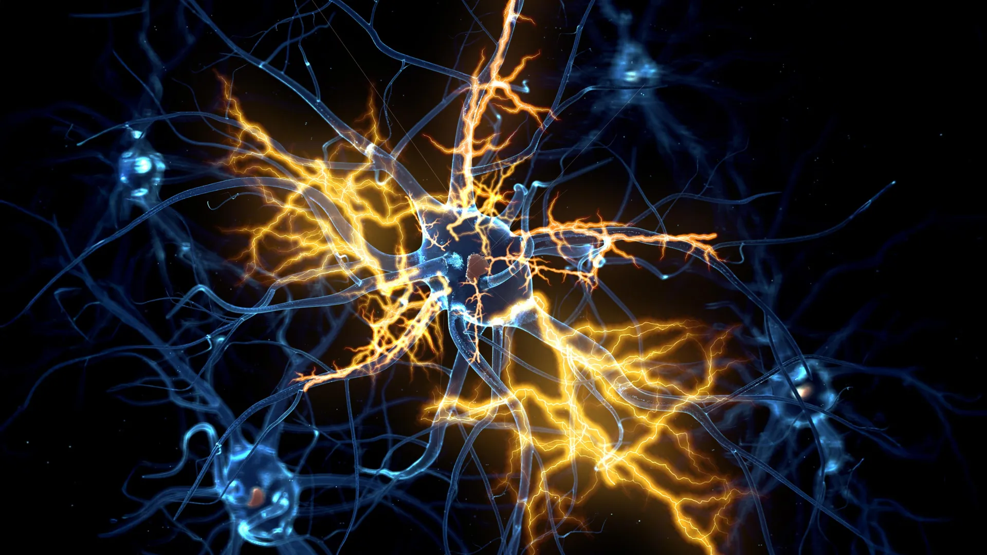
Scientists at the University of Cambridge have engineered sophisticated lab-grown brain and spinal cord systems, providing an unprecedented window into the intricate journey of movement signals through the human nervous system. This groundbreaking model has yielded a remarkable discovery: nerve damage, long considered an irreversible consequence of central nervous system injuries, may indeed be amenable to repair under specific circumstances. The research, published in the esteemed journal Cell Reports, challenges long-held assumptions and injects a potent dose of optimism into the pursuit of treatments for debilitating neurological conditions.
The Intricate Dance of Neural Communication and the Loss of Regeneration
From the earliest stages of embryonic development, the human nervous system orchestrates a symphony of complex communication. Neurons, the fundamental building blocks of this system, extend specialized appendages called axons, which act as conduits for electrical and chemical signals. These signals, originating in the brain and traversing the spinal cord, are responsible for a vast array of functions, most critically, the control of voluntary muscle movement. The precision and speed of this neural network are crucial for everything from a simple gesture to intricate athletic feats.
However, a critical evolutionary shift occurs as the central nervous system matures. The remarkable regenerative capacity that allows developing axons to navigate and establish connections in the embryonic and fetal stages significantly diminishes in adulthood. This inherent limitation means that injuries to the brain or spinal cord, such as those resulting from traumatic accidents or strokes, often lead to permanent functional deficits. Conditions like paralysis, paraplegia, and quadriplegia are stark reminders of this regenerative deficit. Furthermore, this age-related decline in axonal regrowth is implicated in the pathogenesis of progressive neurodegenerative diseases, including amyotrophic lateral sclerosis (ALS), also known as motor neurone disease, and multiple sclerosis (MS). In these conditions, the gradual degeneration of neurons and their axons leads to progressive loss of motor control, sensory function, and cognitive abilities, significantly impacting the quality of life for millions worldwide.
Miniature Human Nervous Systems: A Revolution in Neurological Research
Building upon their prior success in creating pea-sized "brain organoids" from patient-derived stem cells in 2021, Dr. András Lakatos and his team at the University of Cambridge have now advanced their research to an even more complex and revealing level. Their previous work, which focused on modeling aspects of the cerebral cortex to study molecular changes associated with motor neurone disease, laid the groundwork for this latest endeavor. The new study details the creation of a miniature, interconnected human brain and spinal cord system.
To accurately replicate the separated yet functionally linked nature of these structures in the body, the researchers ingeniously cultured the brain and spinal cord organoids physically apart in their laboratory environment. The critical observation was the spontaneous growth of axons from the brain tissue across the interstitial gap to establish connections with the spinal cord tissue. This remarkable display of neural plasticity, even in a lab-grown system, allowed the researchers to witness the formation of functional neural circuits. Crucially, these engineered circuits were robust enough to initiate contractions in attached clusters of muscle cells, effectively mimicking the downstream effects of nerve signaling on motor function. This level of integrated functionality in a simplified model represents a significant leap forward in understanding how these complex systems operate and respond to perturbation.
Unraveling the Developmental Timeline of Regenerative Decline
The Cambridge team meticulously maintained these miniature brain and spinal cord systems in their laboratory for an extended period, exceeding one year. This longitudinal observation allowed them to chart the changes in axonal regenerative capacity over a developmental timeline. Their findings revealed a critical window: up until approximately day 150 of development, a stage roughly corresponding to the second trimester of human gestation, damaged axons within the organoids exhibited a notable ability to regrow. However, beyond this developmental milestone, the neurons displayed a precipitous decline in their regenerative potential.
George Gibbons, a key researcher from the Department of Clinical Neurosciences at the University of Cambridge and the first author of the study, elaborated on these findings. "Neurons taken from less mature organoids regrew long fibers after injury, but those from more mature organoids showed a sharp drop in their ability to regrow," Gibbons stated. "In other words, poor regeneration is built into human neurons as they mature in the central nervous system." This observation suggests that the loss of regenerative capacity is not a random event but a programmed aspect of neural development, intrinsically linked to the maturation process of the central nervous system.
The Genetic Switch Controlling Axonal Growth
Delving deeper into the molecular mechanisms underlying this regenerative decline, the scientists conducted a comprehensive analysis of gene activity in the neurons connecting the brain and spinal cord. Their investigation uncovered a complex network of genes that appears to function as a sophisticated biological "switch." This genetic machinery is responsible for orchestrating the limitation of axon growth as neurons mature and establish synaptic connections, a process essential for efficient information processing but detrimental to repair after injury.
The implications of this discovery are profound. By identifying the specific genetic regulators involved, the researchers opened the door to manipulating these pathways. In a pivotal experiment, when the researchers selectively blocked key regulators within this identified gene network, the mature neurons unexpectedly regained their capacity for axonal growth. This intervention demonstrated that the decline in regeneration is not an immutable fate but a dynamically controlled process that can be reversed.
A Pharmaceutical Ally in Nerve Regeneration: Lynestrenol
The identification of this crucial gene network prompted the researchers to explore existing pharmacological agents that might influence its activity. Leveraging extensive databases of drug compounds, they sought out medicines known to interact with the identified genetic pathways. Among the promising candidates identified was lynestrenol, a synthetic progestogen hormone. Lynestrenol is currently approved and utilized for specific gynecological conditions, including the management of menstrual disorders and as a component of certain contraceptive formulations.
The decision to test lynestrenol on the damaged neurons was driven by its known biological targets, which showed overlap with the newly discovered gene network regulating axon growth. When administered to the lab-grown neurons exhibiting limited regenerative capacity, lynestrenol demonstrated a statistically significant enhancement in axon regrowth. This finding suggests that a drug already in clinical use for other indications could potentially be repurposed to promote nerve repair.
While the current research focused on the intrinsic cellular mechanisms limiting regeneration, the Cambridge team acknowledges the additional challenges posed by the post-injury environment. Scar tissue formation and inflammation are well-documented impediments to nerve repair, often creating physical and biological barriers that prevent axons from regrowing. However, the current study’s emphasis on neuron-specific biological mechanisms provides a crucial complementary perspective. Previous research has indicated that younger neurons possess a greater ability to navigate and grow through environments that would typically inhibit repair in more mature systems. This suggests that by restoring the inherent regenerative capacity of the neurons themselves, the obstacles presented by the surrounding tissue might become more surmountable.
Senior author Dr. András Lakatos underscored the significance of these findings. "When the brain and spinal cord are damaged, the nerve fibers that carry movement signals from the brain to the spinal cord rarely grow back. That’s why paralysis is usually permanent," he stated. "But we didn’t know exactly when the ability of axons to regenerate becomes limited. Our model provides a good indication that this block happens during development, and it can still be reversed after this point." He further elaborated on the potential of lynestrenol: "Lynestrenol itself may not be the answer to spinal cord repair, but it shows us that, in principle, it should be possible to directly target human neurons and regenerate their axons. Although we still need to show that this strategy will also help to re-establish appropriate connections between the brain and spinal cord cells, this gives us hope that one day we may be able to treat conditions previously thought untreatable."
The Growing Importance of Human Organoids in Medical Advancement
The development and application of organoid technology, as exemplified by this research, represent a paradigm shift in biological and medical investigation. While animal models, such as mice and rats, have historically been invaluable tools in understanding human biology and disease, crucial biological differences between species can limit the direct translatability of findings to human patients. The nervous systems of rodents, for instance, exhibit distinct regenerative characteristics compared to their human counterparts.
Human stem cell-derived organoids, such as those developed by the Cambridge team, offer a more accurate and relevant biological platform for studying human physiology and pathology. They provide a means to bridge the gap between findings in animal studies and the prediction of real-world patient outcomes. This approach allows researchers to investigate complex human-specific biological processes in a controlled laboratory setting, accelerating the pace of discovery and the development of targeted therapies.
Dr. Lakatos highlighted the value of these models: "Much of what we know about nerve regeneration comes from rodents, whose neurons behave differently from human neurons. Our sophisticated organoid models help bridge the knowledge gap from animal models to what we see in patients. They are also an important contribution to efforts to reduce the use of animals in research." This sentiment aligns with the growing global emphasis on the "3Rs" principle in animal research: Replacement, Reduction, and Refinement.
The University of Cambridge is at the forefront of utilizing organoid technology for a diverse array of medical research endeavors. Beyond neuroregeneration, these innovative models are being employed to study liver regeneration, investigate the complex mechanisms of Crohn’s disease in pediatric patients, and explore the earliest, most delicate stages of human pregnancy. This broad application underscores the versatility and transformative potential of organoid technology across multiple fields of medical science.
The research underpinning this breakthrough was generously funded by the UK Research and Innovation Medical Research Council and Spinal Research, organizations dedicated to advancing medical knowledge and finding solutions for spinal cord injuries. This collaborative effort highlights the critical role of public and private funding in driving pioneering scientific discovery and translating fundamental research into tangible hope for improved human health. The implications of this Cambridge study extend far beyond the laboratory, offering a beacon of possibility for individuals affected by currently untreatable neurological conditions.
