
 By Nana O
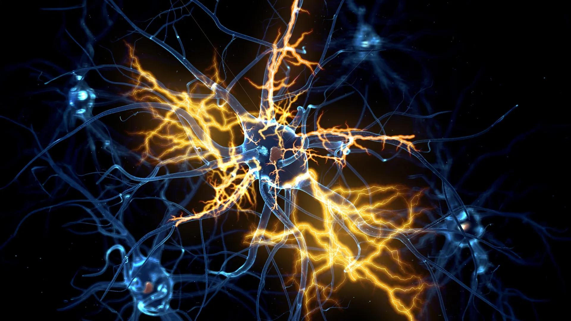
Scientists at the University of Cambridge have achieved a significant breakthrough in neuroscience by developing sophisticated lab-grown brain and spinal cord systems that meticulously replicate the intricate pathways of movement signaling in the human nervous system. This groundbreaking model has led to a paradigm shift in our understanding of nerve damage, revealing that injuries previously considered irreversible may, in fact, be amenable to repair under specific conditions. The findings, published in the esteemed journal Cell Reports, offer a beacon of hope for individuals suffering from debilitating neurological conditions.
The Intricate Dance of Neural Communication and the Challenge of Regeneration
The journey of human development is intrinsically linked to the formation of complex neural networks. From the embryonic stage through fetal development and into infancy, specialized cells called neurons establish intricate communication lines between the brain and the spinal cord. These messages, crucial for controlling voluntary movement and sensory perception, are transmitted via axons – the long, slender extensions of neurons that act as conduits for electrochemical signals. The precise and rapid transmission of these signals is paramount for everything from simple reflexes to complex motor skills.
However, a fundamental challenge arises as the central nervous system matures. The remarkable regenerative capacity observed in developing neurons largely diminishes in adulthood. This loss of ability to regrow damaged axons means that injuries to the brain or spinal cord, such as those sustained in accidents or due to degenerative diseases, often result in permanent functional deficits. Conditions like paralysis, loss of motor control, and sensory impairments are grim realities for millions worldwide. Furthermore, this regenerative decline is a significant factor in the progression of devastating neurological disorders, including motor neurone disease (also known as Amyotrophic Lateral Sclerosis or ALS) and multiple sclerosis, where the progressive loss of myelin sheaths and neuronal damage leads to increasing disability.
Pioneering Miniature Human Nervous Systems
Building upon their previous success in creating pea-sized "brain organoids" from patient-derived stem cells in 2021, a team led by Dr. András Lakatos at the University of Cambridge has now engineered a more comprehensive model: a miniature, interconnected human brain and spinal cord system. These organoids, designed to mimic specific regions of the cerebral cortex and spinal cord, allow researchers to delve into the molecular mechanisms underlying neurological diseases and explore potential therapeutic interventions with unprecedented fidelity.
The innovative approach involved keeping the brain and spinal cord organoids physically separate in the laboratory environment, acknowledging their distinct yet interconnected roles within the body. Crucially, the researchers observed axons extending from the brain tissue successfully bridging the gap and forming functional connections with the spinal cord tissue. This remarkable feat of bioengineering resulted in a nascent neural circuit capable of eliciting contractions in specially cultured clusters of muscle cells, demonstrating the model’s ability to recapitulate fundamental aspects of motor control signaling.
A Developmental Clock for Nerve Regrowth
The Cambridge team meticulously monitored these miniature neural systems in vitro for over a year, observing their developmental trajectory and regenerative capabilities. Their findings revealed a critical developmental window: up until approximately day 150 of development, a period roughly corresponding to the middle trimester of human gestation, damaged axons within these nascent systems exhibited a notable capacity for regrowth. Beyond this point, however, the neurons displayed a dramatic and significant decline in their regenerative potential.
George Gibbons, a researcher from the Department of Clinical Neurosciences at the University of Cambridge and the study’s lead author, elaborated on this pivotal discovery: "Neurons derived from less mature organoids demonstrated the ability to regrow extensive fibers following injury. In stark contrast, those originating from more mature organoids showed a precipitous drop in their regenerative capacity. This strongly suggests that a diminished ability to regenerate is an inherent characteristic that develops within human neurons as they mature within the central nervous system."
Unraveling the Genetic Switch for Regeneration
To understand the biological underpinnings of this developmental decline, the researchers undertook a comprehensive analysis of gene activity within the neurons connecting the brain and spinal cord. Their investigation identified a complex network of genes that appears to function as a sophisticated biological switch. This genetic circuitry plays a pivotal role in regulating and ultimately limiting axon growth as neurons mature and establish synaptic connections.
In a striking demonstration of this mechanism, the scientists were able to experimentally block key regulatory elements within this identified gene network. The outcome was remarkable: the mature neurons, previously exhibiting limited regenerative capacity, regained their ability to grow axons once more. This finding provides compelling evidence that the observed decline in regeneration is not an immutable biological fate but rather a tightly regulated process that can potentially be modulated.
A Pharmaceutical Ally in Nerve Repair
The identification of this crucial gene network opened up new avenues for therapeutic exploration. The Cambridge team embarked on a search through a comprehensive database of drug compounds, seeking existing medications that might interact with and influence this newly discovered genetic circuitry. Among the promising candidates identified was lynestrenol, a hormone-based drug with established therapeutic uses in managing certain menstrual disorders and serving as a contraceptive.
When lynestrenol was administered to damaged neurons in the laboratory setting, the results were highly encouraging. The drug demonstrated a significant enhancement in axon regrowth, offering tangible proof that pharmacological interventions could potentially restore lost regenerative capabilities.
While acknowledging that scar tissue and inflammation can present additional hurdles to nerve repair following injury, the researchers emphasize the paramount importance of understanding the neuron-specific biological mechanisms that inherently limit regeneration. Previous research has indicated that younger, more adaptable neurons can often navigate and grow through environments that would typically impede repair in more mature nervous systems.
Dr. András Lakatos, the senior author of the study and leader of the research group, underscored the significance of these findings: "When the brain and spinal cord are subjected to damage, the nerve fibers responsible for transmitting movement signals from the brain to the spinal cord have a very low propensity to regrow. This is the primary reason why paralysis is typically a permanent condition. However, our research has now provided a clearer understanding of precisely when the capacity for axon regeneration becomes limited. Our organoid model offers compelling evidence that this block develops during the maturation process, and crucially, that it can potentially be reversed even after this developmental point."
He further articulated the broader implications: "While lynestrenol itself may not be the ultimate solution for spinal cord repair, its efficacy demonstrates a fundamental principle: it is potentially possible to directly target human neurons and stimulate the regeneration of their axons. Although further research is necessary to confirm whether this strategy can also facilitate the re-establishment of appropriate functional connections between brain and spinal cord cells, this discovery ignites hope that conditions once deemed untreatable might one day become amenable to effective therapies."
The Indispensable Role of Human Organoids in Advancing Neuroscience
The burgeoning field of organoid technology is proving to be an invaluable asset in unraveling the complexities of human biology and disease. While animal models, such as rodents, have historically played a crucial role in scientific research, inherent biological differences between species can limit their predictive accuracy when it comes to human nervous system function.
Human stem cell-derived organoids offer a more faithful recapitulation of human biology, effectively bridging the gap between preclinical animal studies and the real-world outcomes observed in patients. This enhanced biological relevance allows for more precise investigation of human-specific disease mechanisms and the development of more targeted and effective treatments.
Dr. Lakatos highlighted the comparative advantage of human organoids: "A significant portion of our current knowledge regarding nerve regeneration has been derived from studies on rodents, whose neurons exhibit distinct behavioral patterns compared to human neurons. Our sophisticated organoid models are instrumental in closing this knowledge gap, translating findings from animal models to a more accurate representation of what we observe in human patients. Furthermore, these models represent a significant contribution to the ongoing global efforts aimed at reducing the reliance on animal testing in scientific research."
The University of Cambridge is at the forefront of utilizing organoid technology across a diverse spectrum of medical research. Beyond neurosciences, these miniature biological systems are being employed to study liver regeneration, investigate the pathogenesis of Crohn’s disease in pediatric populations, and explore the earliest, most delicate stages of human pregnancy.
The research leading to these significant findings was generously funded by the UK Research and Innovation Medical Research Council and Spinal Research, underscoring the collaborative and well-supported nature of this critical scientific endeavor. The implications of this work are profound, offering a renewed sense of optimism in the pursuit of effective treatments for conditions that have long challenged the medical community.
