
 By George Ongkojoyo
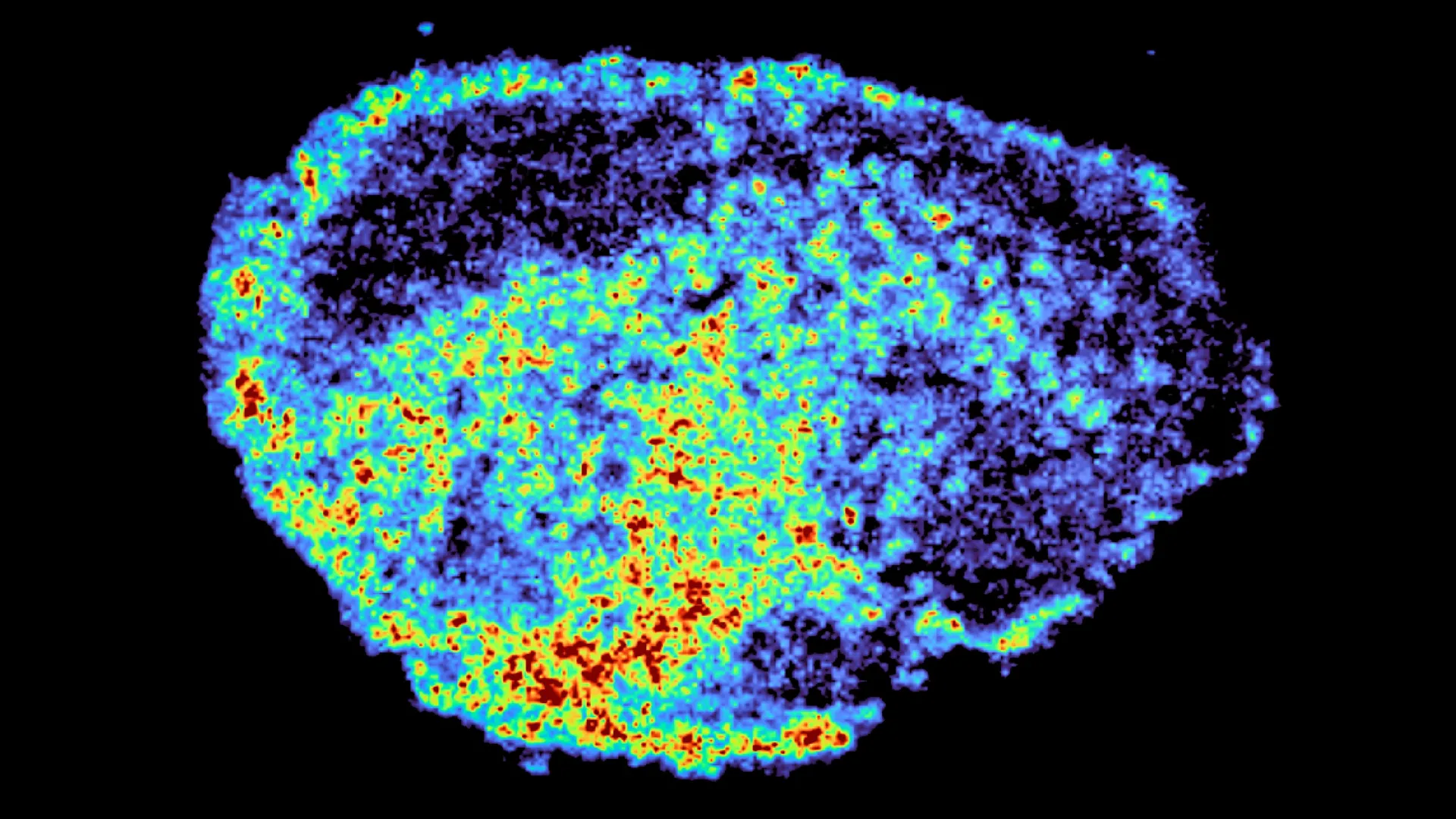
The field of oncology has long grappled with the confounding reality that two patients with seemingly identical diagnoses can respond to the same therapeutic intervention in diametrically opposite ways. While one patient may experience a dramatic reduction in tumor volume, another may see no benefit at all, or worse, develop rapid resistance to the treatment. A groundbreaking study published in the journal Nature Communications has shed new light on this phenomenon, identifying a previously overlooked cellular mechanism that dictates how targeted therapies are distributed and retained within cancerous tissue. Led by Dr. Louise Fets at the MRC Laboratory of Medical Sciences (LMS), the research team utilized cutting-edge imaging technologies to track the movement of PARP inhibitors—a cornerstone of modern ovarian cancer treatment—revealing that these drugs often become sequestered within cellular structures known as lysosomes.
This discovery challenges the traditional understanding of drug efficacy, which has historically focused on whether a drug reaches the tumor via the bloodstream. The new data suggests that the "last mile" of drug delivery—how a molecule maneuvers within the dense architecture of a tumor and the internal compartments of a single cell—is just as critical. By acting as "recycling centers" that double as storage reservoirs, lysosomes can trap certain drugs, creating internal pockets of high concentration while leaving other areas under-treated. This uneven distribution may be a primary driver of treatment failure and the eventual emergence of drug-resistant cancer cell populations.
The Evolution of PARP Inhibitors: A Clinical Context
To understand the significance of this study, one must look at the transformative role PARP inhibitors have played in gynecological oncology over the last decade. Poly (ADP-ribose) polymerase (PARP) is a family of proteins involved in a number of cellular processes, most notably DNA repair. In the context of cancer, PARP inhibitors work by preventing cancer cells from repairing their damaged DNA, eventually leading to cell death. This is particularly effective in tumors with BRCA1 or BRCA2 mutations, which already have a compromised DNA repair mechanism—a concept known as "synthetic lethality."
The first PARP inhibitor, olaparib, received FDA approval in 2014, followed by rucaparib in 2016 and niraparib in 2017. These drugs have significantly extended progression-free survival for thousands of women with ovarian cancer, which remains the most lethal gynecological malignancy in the Western world. Despite these successes, the clinical reality is that many patients eventually relapse. Statistics indicate that while initial response rates to PARP inhibitors can be as high as 60-70% in specific genetic subgroups, the majority of patients with advanced disease will see their cancer return within two to three years. Until now, the mechanisms behind this acquired resistance were thought to be purely genetic, such as "reversion mutations" that restore DNA repair functions. The research from MRC LMS suggests that the physical and chemical distribution of the drug itself is a major, yet understudied, piece of the puzzle.
Methodological Breakthrough: Explants and Spatial Mapping
A central challenge in pharmacological research is the difficulty of observing how a drug behaves inside a living human tumor. Traditional methods often involve grinding up tissue samples to measure average drug concentrations, a process that destroys all spatial information. To overcome this, Dr. Fets and her team employed an innovative "explant" model. They took thin slices of ovarian tumors donated by patients and maintained them in a viable state within a laboratory setting. This allowed the researchers to treat the tissue with PARP inhibitors and observe the drug’s movement through authentic human tumor architecture in real-time.
The team utilized Mass Spectrometry Imaging (MSI), a sophisticated tool that allows scientists to visualize the distribution of specific molecules across a tissue surface with high precision. By firing a laser at the tissue slices, they could identify the chemical signature of the PARP inhibitors and map exactly where they were accumulating. This was then paired with spatial transcriptomics—a method used to measure gene activity in specific locations. By overlaying the drug maps with gene expression maps, the researchers could compare the biological behavior of cells in "drug-rich" zones versus "drug-poor" zones within the same tumor sample.
Dr. Zoe Hall, a senior author of the study and Associate Professor at Imperial College London’s Department of Metabolism, Digestion and Reproduction, emphasized the novelty of this approach. "Through the spatial mapping of drug molecules, we could pinpoint regions of high and low drug and compare gene expression from the same tissue slice. This allowed us to see not just where the drug was, but what the cells were doing in response to those specific concentrations," she noted.
The Lysosomal Mechanism: A Hidden Storage System
The most striking finding of the study was the role of lysosomes. These organelles are typically viewed as the cell’s waste disposal system, filled with acidic enzymes that break down proteins, lipids, and cellular debris. However, the researchers discovered that for certain PARP inhibitors, the lysosomes act as "ion traps." Because some of these drugs are chemically basic, they are drawn into the highly acidic environment of the lysosome. Once inside, they become "protonated"—gaining a positive charge—which prevents them from passing back through the lysosomal membrane.
This sequestration creates a "reservoir effect." The drugs are not immediately available to reach their intended target (the cell nucleus, where DNA repair occurs), but they are also not cleared from the cell. Instead, they are slowly released back into the cytoplasm over time. While this can sometimes be beneficial by prolonging the cell’s exposure to the drug, it also creates massive variability. In some cells, the lysosomes were packed with the drug, while neighboring cells remained virtually untouched.
"We were surprised to see large variability in drug accumulation at the single-cell level," said Dr. Carmen Ramirez Moncayo, the study’s first author and a Postdoctoral Researcher at the LMS. "This variability was driven by the build-up of a drug in lysosomes, which are acting as reservoirs, increasing the exposure of cancer cells to drugs by storing and releasing the drug when needed. However, if the drug is trapped too effectively, it might never reach the nucleus in high enough concentrations to be lethal."
Chemical Divergence: Why Not All PARP Inhibitors Are Equal
A critical takeaway for clinicians is that the lysosomal trapping effect is not universal across all drugs in the PARP inhibitor class. The study specifically highlighted that rucaparib and niraparib—two commonly prescribed treatments—are highly susceptible to being sequestered in lysosomes due to their chemical structure. In contrast, olaparib, the first drug of its kind to be approved, did not show the same tendency to accumulate in these compartments.
This chemical divergence could explain why some patients might fail on one PARP inhibitor but respond to another. It also suggests that the "physicochemical" properties of a drug—its size, charge, and solubility—are just as important as its biological target. This data provides a roadmap for pharmaceutical companies to design next-generation inhibitors that can either bypass the lysosome entirely or exploit it as a controlled-release mechanism.
Expert Commentary and Institutional Collaboration
The research was a collaborative effort involving the MRC Laboratory of Medical Sciences, Imperial College London, and several other leading institutions. The multidisciplinary nature of the team—combining expertise in pharmacology, imaging, and oncology—was essential for interpreting the complex data sets generated by mass spectrometry and spatial transcriptomics.
Dr. Louise Fets, the lead author and Head of the LMS’ Drug Transport and Tumour Metabolism Group, believes this research is a vital step toward truly personalized oncology. "By understanding how drugs are taken up into cells, we can understand whether this influences why cancer drugs work for some people and not for others," Fets explained. "Eventually, we hope to be able to study the molecular signature of a patient’s tumor—including its lysosomal density and pH levels—to help tailor therapeutic approaches in a more personalized way."
External observers in the oncology community have noted that this research addresses a "blind spot" in clinical trials. Most trials focus on the genetic profile of the tumor (the "target"), but few account for the "pharmacokinetic" profile (how the drug moves through the tissue). This study provides the evidence needed to start including spatial drug distribution as a metric in future clinical evaluations.
Broader Clinical Implications and the Road to Personalized Oncology
The implications of this study extend far beyond ovarian cancer. PARP inhibitors are currently being utilized or tested for breast, prostate, and pancreatic cancers, particularly those associated with hereditary mutations. If lysosomal trapping is a widespread phenomenon, it could be a factor in treatment resistance across multiple cancer types.
Furthermore, the discovery of lysosomal reservoirs may lead to the development of "adjunct therapies." For example, drugs that alter lysosomal pH could potentially be used in combination with PARP inhibitors to "unlock" the trapped medication, ensuring a more even and potent distribution throughout the tumor. This strategy, known as lysosomal modulation, is already being explored in other areas of medicine and could now find a critical application in oncology.
The study also highlights the importance of the tumor microenvironment. In a living patient, tumors are not just clusters of cancer cells; they are complex ecosystems containing blood vessels, immune cells, and structural proteins. The researchers noted that in real-world clinical settings, the disorganized nature of tumor vasculature likely exacerbates the uneven drug distribution seen in their lab-grown explants. If a drug is already struggling to reach a certain area of a tumor due to poor blood flow, the added barrier of lysosomal trapping in the cells it does reach could create "refuge zones" where cancer cells can survive and mutate.
Future Research Directions and Funding
While the findings are a significant leap forward, the researchers emphasize that there is more work to be done. The next phase of the project will involve moving from tissue explants to animal models to see how the lysosomal reservoir effect interacts with a functioning circulatory system. The team also plans to study larger cohorts of patients, particularly those who have already relapsed, to see if increased lysosomal trapping is a hallmark of advanced, treatment-resistant disease.
The study was made possible through extensive support from the global scientific community. Funding was provided by the Medical Research Council (MRC) and Cancer Research UK, along with a PhD studentship from the Integrative Toxicology Training Partnership. Significant support also came from the Victoria’s Secret Global Fund for Women’s Cancers Career Development Award, in partnership with Pelotonia and the American Association for Cancer Research (AACR).
As the medical community moves toward an era of "precision medicine," studies like this underscore that precision must apply not only to the genetic target but also to the physical journey of the drug. By mapping the micro-landscape of the tumor and understanding the hidden reservoirs within the cell, scientists are closing the gap between a promising therapy and a permanent cure. The identification of lysosomes as a key factor in drug distribution marks a turning point in the effort to ensure that the right drug reaches the right part of the cell at the right time.
