
 By Budi Oetomo Narendra
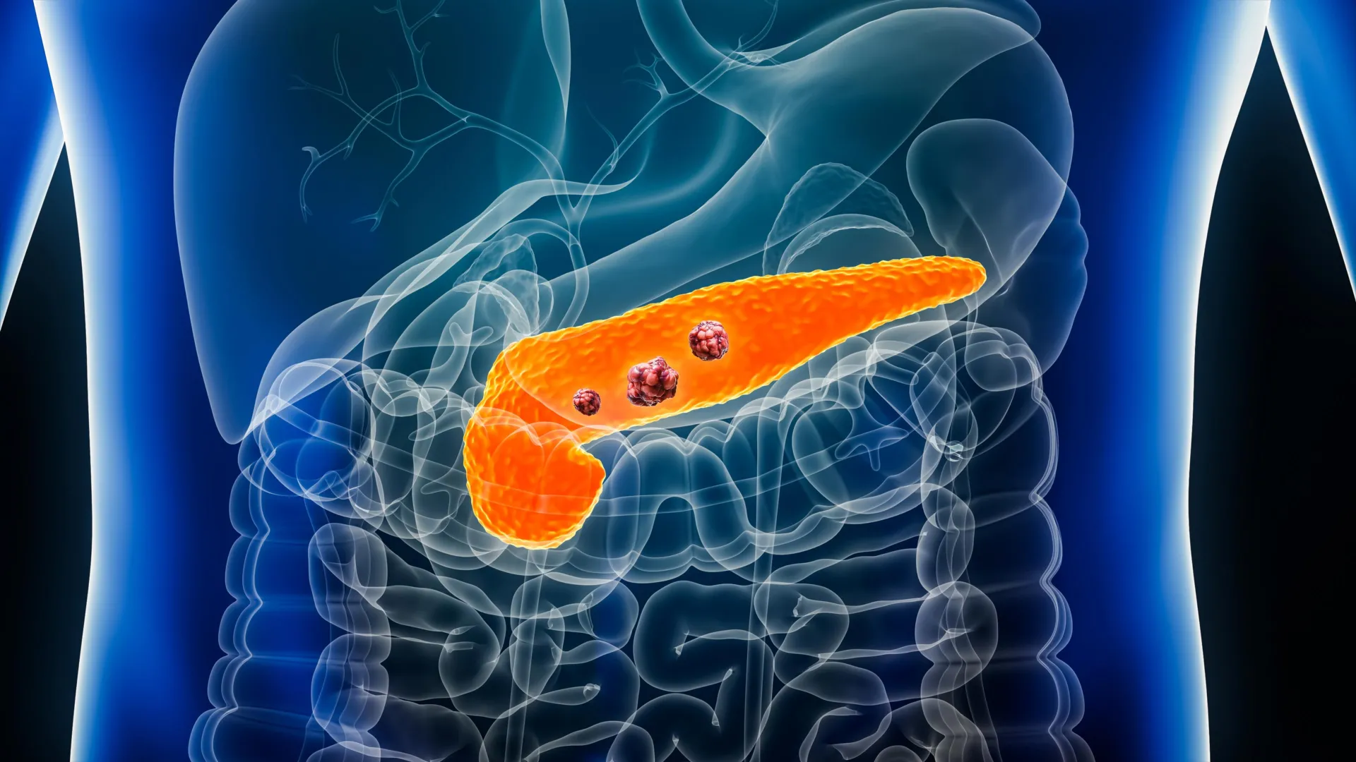
A groundbreaking artificial intelligence model, designated REDMOD (Radiomics-based Early Detection MODel), has demonstrated an unprecedented ability to identify extremely subtle, pre-clinical changes in pancreatic tissue that may signal cancer long before the disease becomes apparent to medical professionals or conventional imaging technologies. This significant development, detailed in research published online in the prestigious journal Gut, marks a potential paradigm shift in the early detection of pancreatic ductal adenocarcinoma (PDAC), the most prevalent and aggressive form of pancreatic cancer. The system’s capacity to discern nascent signs of PDAC offers a critical window for intervention, presenting a new frontier in the battle against a notoriously intractable disease.
Researchers believe this innovative technology holds the promise of revolutionizing pancreatic cancer detection by identifying the disease at its earliest possible stage, often referred to as stage 0. At this nascent phase, when the malignancy is confined and localized, treatment modalities are significantly more likely to achieve curative outcomes. This starkly contrasts with the current reality, where most patients receive a diagnosis only after the cancer has advanced to a more aggressive, often metastatic, and potentially terminal stage. The advent of REDMOD could empower patients with the opportunity to commence treatment while the disease remains localized and highly treatable, fundamentally altering the trajectory of their prognosis.
While the study revealed REDMOD’s superior performance compared to experienced radiologists in detecting these elusive early cancer signals, the scientific community emphasizes the necessity for further rigorous investigation before the model can be seamlessly integrated into routine clinical practice. Specifically, the research team underscores the critical importance of subjecting the system to extensive prospective testing in high-risk patient populations. This includes individuals presenting with unexplained weight loss, new-onset diabetes without obvious cause, or those with a family history of pancreatic cancer, all of whom are considered at elevated risk for developing the disease.
The Formidable Challenge of Early Pancreatic Cancer Detection
Pancreatic ductal adenocarcinoma is infamous for its exceptionally low survival rate, a grim statistic largely attributable to its late-stage discovery. Globally, pancreatic cancer is the seventh leading cause of cancer-related deaths, with its incidence and mortality rates remaining stubbornly high compared to many other cancers. In the United States, for instance, the American Cancer Society estimates that over 66,000 new cases of pancreatic cancer will be diagnosed in 2024, leading to more than 51,000 deaths. The five-year survival rate for PDAC hovers around a mere 12%, a figure that drops significantly for metastatic disease but rises substantially to 44% if detected at a localized stage. This dramatic difference underscores the profound impact early detection could have.
The inherent difficulty in detecting early pancreatic cancer stems from several biological and diagnostic challenges. In its nascent stages, PDAC typically manifests with few, if any, noticeable symptoms. When symptoms do emerge, they are often vague and non-specific, such as abdominal pain, back pain, jaundice, unexplained weight loss, or new-onset diabetes, which can easily be attributed to other less serious conditions. Furthermore, the subtle cellular and architectural changes induced by early-stage cancer within the pancreatic tissue are often imperceptible through standard medical imaging techniques like conventional computed tomography (CT) scans, magnetic resonance imaging (MRI), or ultrasound. Compounding these issues is the aggressive biological nature of PDAC; it can progress rapidly from an early, localized lesion to an advanced, invasive malignancy within a relatively short timeframe, rendering timely diagnosis critically important.
To address these multifaceted challenges, researchers developed REDMOD, an advanced artificial intelligence framework rooted in the principles of radiomics. Radiomics is an emerging field that extracts a vast array of quantitative features from medical images using advanced algorithms, going far beyond what the human eye can discern. These features describe the texture, shape, intensity, and other characteristics of tissues, revealing patterns that may correlate with underlying pathological processes, including early cancer development. REDMOD leverages this capability by analyzing extremely fine, high-dimensional patterns in the texture of pancreatic tissue, known as radiomic features, which are not readily visible or interpretable on conventional CT scans by human observers.
A key innovation of REDMOD lies in its automated identification and outlining of the pancreas. This sophisticated process precisely segments the organ, meticulously separating it from adjacent organs and tissues within the abdominal cavity. This automated segmentation eliminates the need for specialists, such as radiologists or oncologists, to manually trace the organ’s boundaries – a time-consuming and often inconsistent task. Manual outlining is prone to inter-observer variability, meaning different clinicians might draw slightly different borders, leading to inconsistencies in measurement and interpretation. By automating this crucial step, REDMOD significantly reduces these inconsistencies, enhancing the reproducibility and reliability of its analyses.
Unveiling Hidden Cancer Signatures Years Before Clinical Diagnosis
To rigorously evaluate the performance and efficacy of the REDMOD system, the research team conducted a comprehensive retrospective study involving abdominal CT scans from 219 patients. All these patients were subsequently diagnosed with pancreatic cancer after their initial scans, which had, at the time, been deemed clear of any malignant findings by experienced radiologists during routine clinical assessment. The patient cohort was drawn from multiple hospitals, ensuring a degree of external validity to the findings.
The researchers meticulously analyzed the timeline of these scans relative to the eventual clinical diagnosis. For 87 patients, representing 40% of the cohort, the CT images were acquired between 3 and 12 months prior to their pancreatic cancer diagnosis. Another substantial group of 76 patients (35%) had undergone scans between 12 and 24 months before receiving their diagnosis. Remarkably, 56 patients (25%) had imaging performed more than 24 months prior to diagnosis, with some scans taken up to approximately three years in advance. The anatomical distribution of the cancer was also noted, with nearly two-thirds (64%) of the patients developing cancer in the head of the pancreas, a common site for PDAC.
To provide a robust control for comparison, these diagnostic images were contrasted with CT scans obtained from 1,243 individuals who remained free of pancreatic cancer for a follow-up period of up to three years after their scans. This large comparison group was carefully matched to the patient cohort based on critical demographic factors such as age, sex, and the specific dates of their CT scans, minimizing confounding variables and strengthening the statistical validity of the findings.
The demographic analysis revealed that patients who eventually developed pancreatic cancer had an average age of 69 years, with ages spanning a wide range from 34 to 88 years. The individuals in the cancer-free comparison group had an average age of 64 years, exhibiting the same broad age range of 34 to 88 years.
The core finding of the study was REDMOD’s exceptional ability to successfully identify subtle imaging signatures unequivocally associated with pancreatic cancer. Critically, these detections occurred an average of 475 days (approximately 1 year and 3 months) before patients received their official clinical diagnoses. These crucial early warning signs, residing within the complex radiomic patterns of the CT scans, had consistently eluded detection during routine radiological assessments performed by highly trained human experts.
The profound significance of this "temporal window" was emphasized by the researchers. They highlighted that achieving such an early detection capability would "substantially augment the probability of cure and improved survival" for pancreatic cancer patients. They further elaborated, citing modeling studies, that "increasing the proportion of localized [pancreatic ductal carcinomas] from 10% to 50% would more than double survival rates." This powerful statement underscores a fundamental truth about cancer outcomes: the timing of diagnosis is arguably the single most critical determinant of survival outcomes for PDAC, often outweighing other factors like specific treatment regimens once the disease has progressed.
REDMOD’s Superiority Over Experienced Radiologists
Beyond merely identifying early signals, the study’s results unequivocally demonstrated REDMOD’s substantial superiority over experienced human radiologists in recognizing these nascent cancer indicators. This comparison is particularly vital for evaluating the practical utility of an AI system in a clinical setting.
The AI model achieved a remarkable sensitivity of 73%. Sensitivity, a key metric in diagnostic testing, quantifies how effectively a diagnostic method correctly identifies individuals who genuinely have the disease, minimizing the number of false negatives (overlooking actual cases). In stark contrast, the experienced radiologists achieved a sensitivity of only 39%. This difference highlights a significant advantage for REDMOD in its ability to avoid missing true positive cases, a critical factor in early cancer detection.
The performance disparity became even more pronounced when the researchers focused their analysis on scans taken more than two years before patients received their official diagnoses. In these particularly challenging cases, where the cancer signals were presumably at their most subtle and elusive, REDMOD achieved an impressive detection rate of 68%. This figure was nearly three times higher than the 23% detection rate recorded by the human radiologists, underscoring the AI’s capability to detect extremely subtle changes that are beyond the perceptive capacity of even highly trained human eyes.
The researchers also rigorously assessed REDMOD’s specificity – its ability to correctly identify scans from individuals who did not have pancreatic cancer, thus minimizing false positives. In an independent validation group comprising 539 patients from several different hospitals, REDMOD correctly classified just over 81% of scans as cancer-free. Its performance was even more robust on the publicly available US National Institutes of Health (NIH-PCT) dataset, which included 80 patients, where it correctly identified 87.5% of scans as free of pancreatic cancer. This high specificity is crucial to avoid unnecessary follow-up procedures, patient anxiety, and healthcare costs associated with false positive results.
Further testing reinforced the reliability of REDMOD’s findings. The early warning signs detected by the AI system demonstrated remarkable consistency over time. When researchers analyzed serial CT scans taken from the same patients several months apart, the system produced matching results in an impressive 90% to 92% of cases. This high degree of consistency strengthens the evidence that REDMOD is indeed identifying meaningful, stable changes associated with the very early stages of pancreatic cancer, rather than random fluctuations or imaging artifacts.
Could AI Transform Early Pancreatic Cancer Detection? Implications and Future Directions
Despite these highly promising findings, the researchers prudently caution that several critical questions remain unanswered and necessitate further investigation. One acknowledged limitation of the study is the demographic composition of the study population, which did not include a sufficiently diverse range of ethnic backgrounds. This lack of diversity could potentially impact how effectively the technology performs across different populations, given that variations in genetics, lifestyle, and disease presentation can exist across ethnic groups. Addressing this requires broader, multi-center studies with more diverse cohorts.
Furthermore, the system’s true clinical utility and effectiveness in everyday medical practice hinge upon robust prospective testing. This involves deploying REDMOD in real-time clinical scenarios, particularly among individuals identified as being at an increased risk of pancreatic cancer, and observing its performance in predicting future diagnoses. Such prospective validation is paramount for securing regulatory approvals, such as from the U.S. Food and Drug Administration (FDA), and for gaining widespread acceptance within the medical community.
Even with these acknowledged limitations and the need for further validation, the findings from the REDMOD study represent a monumental step forward. They strongly suggest that advanced AI could eventually equip doctors with the tools to identify pancreatic cancer while it is still at a much earlier, significantly more treatable stage. This potential shift holds immense promise for dramatically improving survival rates and overall outcomes for patients who, under current diagnostic paradigms, would regrettably receive a late-stage diagnosis with a far grimmer prognosis.
The integration of such AI technologies into healthcare carries broader implications. For healthcare systems, it could lead to more efficient allocation of resources by reducing the need for expensive, aggressive treatments for advanced cancers. For public health, it could pave the way for novel screening strategies, potentially targeting high-risk groups with non-invasive AI-powered assessments. However, challenges related to the cost-effectiveness of AI implementation, data privacy, and the ethical considerations of AI in diagnostic decision-making will need to be carefully navigated. Education and training for radiologists and oncologists will also be crucial to ensure seamless adoption and effective utilization of these advanced tools.
In their concluding remarks, the researchers articulated the profound significance of their work: "This study validates REDMOD as a fully automated AI framework capable of identifying the imaging signatures of stage 0 [pancreatic ductal adenocarcinoma] in normal pancreas, achieving this with substantial lead times and performance superior to expert radiologists." They further emphasized the transformative potential, adding, "While prospective validation is paramount to confirm clinical utility, the REDMOD framework represents a significant advance towards shifting the paradigm for sporadic [pancreatic ductal adenocarcinoma] from a late-stage symptomatic diagnosis to proactive pre-clinical interception, offering tangible hope for improving outcomes in this challenging disease." This sentiment encapsulates the optimism surrounding REDMOD, envisioning a future where pancreatic cancer, once a silent killer, may finally be caught in time to be cured.
