
 By George Ongkojoyo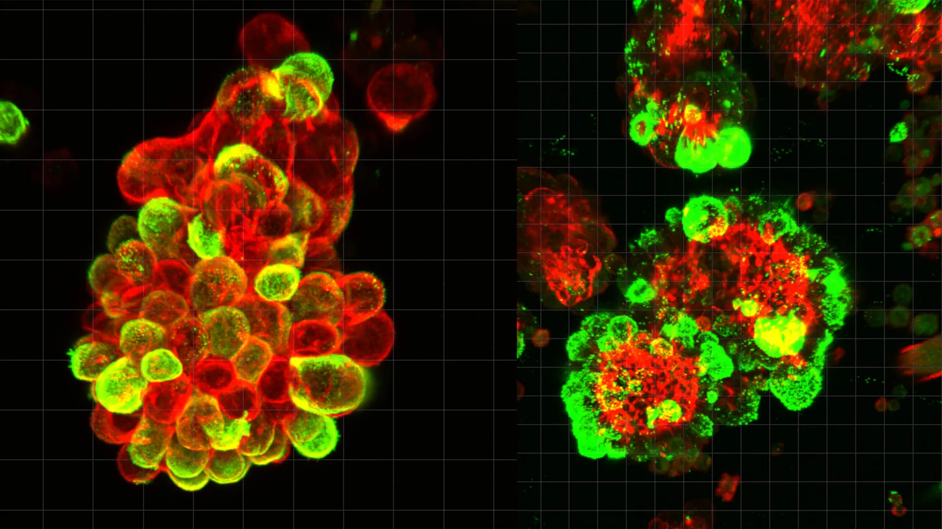
Ovarian cancer remains the most lethal of all gynecological malignancies, a reality driven primarily by its propensity for late-stage detection and its uniquely aggressive method of spreading through the abdominal cavity. While the medical community has long recognized the speed at which this cancer progresses, the underlying biological mechanisms that facilitate such rapid movement have remained a significant scientific blind spot. However, a groundbreaking study led by researchers at Nagoya University in Japan has finally decoded this mystery, revealing that ovarian cancer cells do not act in isolation. Instead, they actively recruit and "reprogram" healthy cells to act as invasive pioneers, paving the way for the cancer to colonize distant organs within the abdomen.
The research, recently published in the prestigious journal Science Advances, identifies a sophisticated partnership between ovarian cancer cells and mesothelial cells. Mesothelial cells typically form a protective, low-friction lining known as the mesothelium, which covers the internal organs and the abdominal cavity. Under normal conditions, these cells serve as a barrier against infection and injury. However, the Nagoya University team discovered that ovarian cancer cells subvert this protective function, transforming mesothelial cells into accomplices that facilitate tissue invasion and provide a shield against standard chemotherapy treatments.
The Mechanism of Hybrid Cell Clusters
To investigate the dynamics of ovarian cancer progression, the research team, led by Dr. Kaname Uno and Professor Hiroaki Kajiyama, meticulously analyzed abdominal fluid—medically known as ascites—collected from patients diagnosed with ovarian cancer. Their findings immediately challenged the long-standing assumption that cancer cells primarily drift through the abdomen as solitary units.
The analysis revealed that cancer cells frequently aggregate with mesothelial cells that have been shed from the abdominal lining. These two distinct cell types bind together to form compact, "hybrid" multicellular spheres. The researchers estimated that approximately 60% of the cancer cell clusters found in patient fluid samples contained these recruited mesothelial cells. This discovery suggests that the formation of hybrid spheres is a standard operating procedure for ovarian cancer, rather than an incidental occurrence.
The catalyst for this transformation is a signaling molecule secreted by the cancer cells: Transforming Growth Factor-beta 1 (TGF-β1). When the cancer cells release TGF-β1, it acts as a molecular command that fundamentally alters the behavior and structure of the attached mesothelial cells. In response to this signal, the mesothelial cells undergo a process that causes them to develop invadopodia—sharp, spike-like cellular protrusions. These invadopodia act as biological "drills," capable of piercing the basement membranes of healthy organs, creating pathways that the cancer cells then exploit to embed themselves and grow.
A Departure from Conventional Metastasis
The study highlights how ovarian cancer differs fundamentally from other common cancers, such as breast or lung cancer, in its mode of dissemination. In most solid tumors, metastasis occurs via the bloodstream or the lymphatic system (hematogenous or lymphatic spread). Cancer cells enter vessels, travel through the circulatory system, and eventually lodge in distant organs like the liver or brain. Because blood flows through a defined and predictable network of vessels, clinicians can often monitor these cancers through blood-based biomarkers or "liquid biopsies."
Ovarian cancer, however, utilizes "transcoelomic spread." When cells detach from the primary tumor in the ovaries or fallopian tubes, they enter the peritoneal fluid that fills the space between abdominal organs. This fluid is in a constant state of flux, driven by the rhythmic motions of the diaphragm during breathing and the peristaltic movements of the digestive tract. This creates a chaotic and unpredictable "floating phase" where cancer cells can be carried to any surface within the abdomen, from the liver and stomach to the intestines and diaphragm.
Until this study, the scientific community lacked a clear understanding of the biological coordination occurring during this floating phase. The Nagoya University findings demonstrate that the floating phase is not merely a passive period of transport but an active stage of "recruitment and preparation," where cancer cells assemble their invasive toolkit by hijacking mesothelial cells.
The Role of Invadopodia in Tissue Invasion
The most alarming aspect of these hybrid spheres is their enhanced ability to penetrate healthy tissue compared to solitary cancer cells. The mesothelial cells, once activated by the cancer’s TGF-β1 signal, essentially take over the "heavy lifting" of the invasion process. By producing invadopodia, they break down the extracellular matrix of the target organ.
"The cancer cells themselves remain relatively unchanged in terms of their invasive potential," explained Dr. Kaname Uno, the study’s lead author. "Instead, they manipulate the mesothelial cells to do the work of tissue invasion. The cancer cells undergo minimal genetic and molecular changes themselves; they simply migrate through the openings that the mesothelial cells create for them."
This "outsourcing" of invasive labor allows the cancer to spread with remarkable efficiency. Furthermore, the study found that these hybrid clusters are significantly more resistant to common chemotherapy agents, such as cisplatin and paclitaxel, than cancer cells that have not recruited mesothelial "bodyguards." The structural arrangement of the hybrid sphere and the molecular changes within the mesothelial cells appear to create a protective microenvironment that prevents drugs from effectively reaching and killing the malignant cells.
Methodology and Real-Time Observation
The research team employed a multi-disciplinary approach to validate their findings. Using advanced live-cell imaging and high-resolution microscopy, they were able to observe the formation and invasive behavior of these hybrid spheres in real-time using fluid samples taken directly from human patients.
To confirm the clinical relevance of these observations, the researchers utilized mouse models of ovarian cancer. By manipulating the TGF-β1 signaling pathway in these models, they were able to demonstrate that blocking the recruitment of mesothelial cells significantly slowed the rate of metastasis and improved the efficacy of chemotherapy. Additionally, the team performed single-cell RNA sequencing to analyze gene activity within the hybrid clusters. This allowed them to map the exact molecular changes that occur when a normal mesothelial cell is transformed into a pro-cancer invasive agent.
The Clinical Inspiration: A Physician’s Perspective
The motivation for this intensive research was deeply personal for Dr. Kaname Uno. Before transitioning into full-time research as a PhD student and Visiting Researcher at Nagoya University’s Graduate School of Medicine, Uno spent eight years practicing as a gynecologist.
During his clinical tenure, he encountered a patient whose case left a lasting impact on his career. The patient had undergone a routine gynecological screening that showed no signs of abnormality. However, just three months later, she returned with symptoms that led to a diagnosis of advanced, Stage IV ovarian cancer. Despite the best available treatments, the disease had progressed too rapidly for medical intervention to be successful.
"The fact that current diagnostic tools failed to detect the disease in such a short window, and the speed at which it overwhelmed her system, convinced me that we needed to understand the ‘why’ behind this rapid spread," Dr. Uno stated. This experience drove him to investigate the specific interactions in the abdominal fluid that allow ovarian cancer to bypass the body’s natural defenses so effectively.
Implications for Future Treatment and Diagnostics
The discovery of the mesothelial-cancer cell partnership opens several new avenues for the management of ovarian cancer. Current treatment protocols are largely "cancer-centric," focusing on killing the malignant cells themselves. However, the Nagoya University study suggests that the "microenvironment"—the healthy cells surrounding the tumor—is just as critical a target.
- Targeted Therapies: Future drugs could be developed to specifically block the TGF-β1 signaling pathway, preventing cancer cells from being able to recruit or reprogram mesothelial cells. By keeping the mesothelial cells in their natural, protective state, doctors might be able to "trap" cancer cells in the floating phase, making them more vulnerable to existing treatments.
- Preventing Adhesion: Therapies aimed at preventing the physical binding of cancer cells to mesothelial cells could stop the formation of hybrid spheres altogether.
- Enhanced Diagnostics: Monitoring the presence and concentration of these hybrid cell clusters in abdominal fluid could serve as a powerful prognostic tool. By analyzing a patient’s ascites, doctors could potentially predict how aggressively the cancer will spread and tailor chemotherapy regimens based on the level of resistance indicated by the cell clusters.
- Overcoming Chemoresistance: If the mechanism by which mesothelial cells protect cancer cells is fully understood, researchers may be able to develop "sensitizing" agents that break down the hybrid sphere’s defenses, allowing standard chemotherapy to work more effectively.
Chronology of Ovarian Cancer Progression
Based on the study’s findings, the timeline of ovarian cancer spread can now be viewed as a four-stage process:
- Stage 1: Detachment. Cancer cells break away from the primary tumor on the ovary or fallopian tube.
- Stage 2: Recruitment. While floating in the peritoneal fluid, cancer cells release TGF-β1 to attract and bind with shed mesothelial cells.
- Stage 3: Transformation. The hybrid spheres form, and the mesothelial cells develop invadopodia, transforming from passive liners into active invaders.
- Stage 4: Colonization. The hybrid spheres land on abdominal organs. The mesothelial "spikes" drill into the tissue, allowing the cancer cells to enter, establish a blood supply, and form secondary tumors.
Conclusion
Ovarian cancer remains a formidable challenge for global health, with the World Health Organization reporting over 300,000 new cases and 200,000 deaths annually. The high mortality rate is inextricably linked to the disease’s "stealth" nature and its rapid colonization of the abdomen.
By identifying the mesothelial cell as a key "accomplice" in this process, the researchers at Nagoya University have provided a new roadmap for intervention. This shift in focus from the cancer cell alone to the "hybrid partnership" represents a significant leap forward in oncology. As researchers work to translate these laboratory findings into clinical trials, there is renewed hope that the "silent killer" may one day be silenced by therapies that disrupt its most effective means of survival.
