
 By George Ongkojoyo
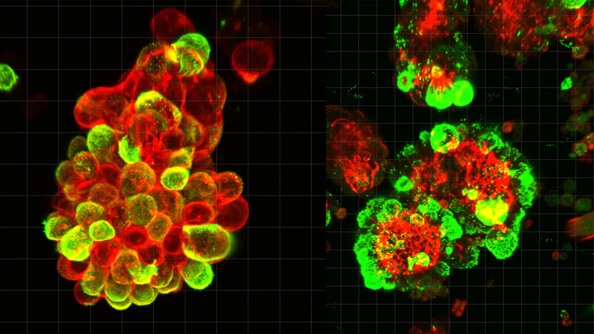
Ovarian cancer remains the most lethal gynecological malignancy worldwide, a grim status primarily attributed to its propensity for late-stage diagnosis and its unique, rapid mode of metastasis within the abdominal cavity. While medical science has long recognized the aggressive nature of this disease, the specific biological mechanisms that allow ovarian cancer cells to colonize the abdomen with such speed and efficiency have remained largely speculative. A landmark study led by researchers at Nagoya University, recently published in the journal Science Advances, has finally unmasked a sophisticated "cooperative" strategy used by cancer cells to hijack healthy tissue. The research reveals that ovarian cancer cells do not act as solitary invaders; instead, they actively recruit and manipulate mesothelial cells—the very cells intended to protect the abdominal lining—to act as biological "drill bits" that pave the way for tumor invasion.
A Paradigm Shift in Understanding Peritoneal Metastasis
For decades, the prevailing understanding of cancer metastasis focused on the "seed and soil" hypothesis, where cancer cells (the seeds) travel through the bloodstream or lymphatic system to find hospitable organs (the soil). In cancers such as breast, lung, or prostate cancer, this hematogenous spread follows relatively predictable vascular routes, allowing for monitoring via blood-based biopsies. Ovarian cancer, however, defies this convention. It primarily spreads through the peritoneal fluid, a lubricating liquid that fills the space between abdominal organs.
The Nagoya University study highlights that during this "floating phase," ovarian cancer cells undergo a transformative partnership. Rather than drifting as individual entities, they aggregate with mesothelial cells—which are naturally shed from the peritoneal lining—to form compact, hybrid cell spheres. These spheres are not merely passive clusters; they are highly active, predatory units. By analyzing abdominal fluid samples from human patients, the research team discovered that approximately 60% of these floating cancer cell clusters contained recruited mesothelial cells. This finding suggests that the formation of hybrid spheres is not a rare occurrence but a fundamental characteristic of ovarian cancer’s progression.
The Molecular Hijacking: TGF-β1 and the Creation of Invadopodia
The mechanism behind this cellular recruitment is a masterclass in biological manipulation. The Nagoya University team identified a specific signaling molecule, Transforming Growth Factor-beta 1 (TGF-β1), which is secreted in high concentrations by the ovarian cancer cells. When mesothelial cells come into contact with this protein, they undergo a profound phenotypic shift. Normally, mesothelial cells provide a smooth, low-friction surface that allows organs to glide against one another. Under the influence of TGF-β1, however, these cells become aggressive and invasive.
The study found that the "hijacked" mesothelial cells begin to produce invadopodia—specialized, actin-rich protrusions that function like microscopic drills. These spike-like structures are capable of secreting enzymes that degrade the extracellular matrix, effectively cutting through the protective layers of the diaphragm, liver, and intestinal walls.
Dr. Kaname Uno, the study’s lead author and a Visiting Researcher at Nagoya University’s Graduate School of Medicine, noted that this discovery changes the way scientists view the "aggressiveness" of cancer cells. "The cancer cells themselves remain relatively passive in terms of physical invasion," Dr. Uno explained. "Instead of undergoing the complex genetic and molecular changes usually required for a cell to become invasive, they simply outsource the hard work. They manipulate the mesothelial cells to do the digging, and then the cancer cells follow through the pathways that have been created for them."
Chronology of the Discovery: From Clinic to Laboratory
The impetus for this research was born from clinical frustration and a desire to address a glaring gap in diagnostic capabilities. Dr. Uno, who served as a practicing gynecologist for eight years before transitioning into full-time research, was motivated by a specific case that highlighted the limitations of modern medicine. One of his patients had undergone a routine gynecological screening that showed no abnormalities. Yet, a mere three months later, she was diagnosed with advanced-stage ovarian cancer that had already metastasized throughout her abdominal cavity.
This rapid progression—occurring in a window of only 90 days—prompted Dr. Uno to investigate the "hidden" stage of ovarian cancer spread: the time spent floating in the peritoneal fluid. The research timeline proceeded through several rigorous phases:
- Clinical Sampling: The team collected and analyzed peritoneal fluid (ascites) from patients at various stages of ovarian cancer.
- Advanced Microscopy: Using high-resolution real-time imaging, the researchers observed the physical interactions between cancer cells and mesothelial cells within the fluid.
- Single-Cell Analysis: The team utilized single-cell RNA sequencing to identify the gene expression profiles of the cells within the hybrid spheres, confirming the role of the TGF-β1 signaling pathway.
- Validation in Vivo: The findings were validated using mouse models, where blocking the recruitment of mesothelial cells significantly slowed the rate of tumor invasion and improved survival outcomes.
Supporting Data: Resistance to Conventional Therapy
Beyond facilitating spread, the hybrid spheres provide a significant survival advantage to the cancer cells during treatment. One of the most critical findings of the study is that these mixed-cell clusters are substantially more resistant to chemotherapy than cancer cells alone.
In laboratory tests, the researchers exposed both solitary ovarian cancer cells and hybrid spheres to standard platinum-based chemotherapy agents. The results showed that the mesothelial cells within the clusters acted as a biological shield, protecting the interior cancer cells from the drugs. This "communal resistance" explains why many ovarian cancer patients initially respond well to chemotherapy—which kills off the easier-to-reach solitary cells—only to suffer a rapid relapse as the hybrid clusters survive and re-colonize the abdomen.
Data from the study indicates that the presence of these hybrid spheres in a patient’s peritoneal fluid is a strong predictor of poor prognosis. The density and composition of these clusters correlate directly with the speed of recurrence, suggesting that the "partnership" between cancer and mesothelial cells is a primary driver of treatment failure.
Broader Implications for Diagnosis and Treatment
The discovery of the hybrid sphere mechanism offers a new roadmap for the future of ovarian cancer management. Currently, the "gold standard" for monitoring cancer—blood tests for biomarkers like CA-125—is often unreliable for early detection or for tracking localized abdominal spread. The Nagoya University research suggests that analyzing the peritoneal fluid for these specific hybrid clusters could provide a much more accurate "liquid biopsy" for ovarian cancer.
Furthermore, the study opens the door to "anti-partnership" therapies. Rather than focusing solely on killing cancer cells, future treatments could aim to:
- Interrupt Signaling: Develop drugs that block the TGF-β1 protein, preventing the cancer cells from communicating with and recruiting mesothelial cells.
- Target Invadopodia: Create inhibitors that prevent the formation of the spike-like protrusions on mesothelial cells, effectively "disarming" the clusters before they can attach to organs.
- Sensitize Clusters: Formulate treatments that break apart the hybrid spheres, making the individual cancer cells more vulnerable to existing chemotherapy.
Analysis: The Future of Gynecological Oncology
The implications of this study extend beyond ovarian cancer, potentially offering insights into other peritoneal cancers, such as gastric or colorectal malignancies, which also utilize the abdominal fluid for spread. By identifying the mesothelial cell as a "willing accomplice" in the metastatic process, the research shifts the focus of oncology from a cell-centric view to a microenvironment-centric view.
"We have spent decades trying to understand what makes a cancer cell ‘evil,’" says one independent commentator in the field of oncology. "This research shows that the ‘evil’ isn’t just in the cancer cell’s DNA, but in its ability to corrupt the healthy cells around it. This is a profound shift in our understanding of the disease’s ecology."
As ovarian cancer continues to claim the lives of over 180,000 women globally each year, the need for breakthroughs in early detection and treatment remains urgent. The work of Dr. Uno and the Nagoya University team provides a critical piece of the puzzle, explaining the "how" and "why" of the disease’s terrifying speed. While clinical applications of these findings may still be several years away, the identification of the hybrid sphere represents a major step toward transforming ovarian cancer from a "silent killer" into a manageable, or even curable, condition.
The study concludes that by targeting the cellular cooperation that defines ovarian cancer, medicine may finally be able to outpace a disease that has, for too long, moved faster than our ability to detect it. The transition from a gynecologist’s clinical observation to a molecular breakthrough serves as a testament to the power of translational medicine in addressing the most challenging frontiers of human health.
