
 By Budi Oetomo Narendra
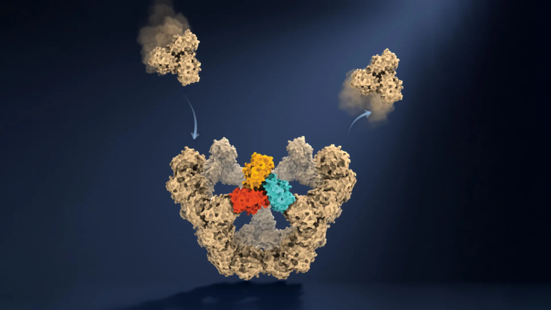
Researchers at the University of Oxford have achieved a groundbreaking feat, directly observing virus-like particles (VLPs) as they meticulously assemble themselves, one molecule at a time. This unprecedented view, detailed in a recent publication in the prestigious journal Nature, has revealed the surprisingly elegant and simple rules governing the formation of complex biological structures with remarkable precision and reliability. The findings offer a long-sought, detailed look into a fundamental biological process that scientists have grappled to observe directly for decades, paving the way for a deeper understanding of viral biology and potential new therapeutic strategies.
A Decades-Long Scientific Challenge Unlocked
For many years, the intricate process by which viruses construct their protective protein shells, known as capsids, has remained a significant enigma. These capsids are essential for safeguarding the virus’s genetic material and facilitating its entry into host cells. Depending on the specific virus, these shells can be composed of tens, hundreds, or even thousands of individual protein components. What makes this process particularly astonishing is that, in many viral systems, these proteins spontaneously organize themselves into the correct, highly ordered structure without the aid of any external machinery or pre-existing blueprint. This phenomenon, termed self-assembly, is a cornerstone of biological organization, yet its real-time dynamics have largely eluded direct observation.
The inherent difficulty in studying viral self-assembly stems from several factors. Many of the intermediate structures that form during the assembly process are incredibly transient, existing only for fleeting moments, and often in minuscule quantities. Traditional biochemical and structural biology methods, such as electron microscopy or X-ray crystallography, typically provide "snapshots" of starting materials and finished particles, or require large, stable quantities of intermediates. This has meant that researchers could infer the process but not directly witness the transformation from individual components to a fully formed, functional capsid. The Oxford team’s innovation lies in overcoming these limitations, providing an unprecedented, continuous "video" of the assembly process.
The Methodology: Seeing the Unseen with Mass Photometry
To circumvent the challenges of observing these ephemeral molecular events, the Oxford researchers employed an engineered virus-like particle (VLP) model, comprising 60 identical protein units designed to self-assemble. The true innovation, however, came from their deployment of a cutting-edge technique known as mass photometry. Developed at the University of Oxford, mass photometry is a revolutionary label-free method that allows scientists to determine the mass of individual molecules in solution by precisely measuring the amount of light they scatter. This highly sensitive technique provides a direct, quantitative measure of molecular weight, making it ideal for tracking the incremental addition of building blocks to a growing structure.
Critically, the team combined mass photometry with a novel approach for confining individual molecules, ensuring they remained continuously observable within the instrument’s field of view. This ingenious combination allowed them to repeatedly measure the mass of a single, growing particle as additional proteins attached, effectively tracking the construction process as it unfolded in real-time. This continuous monitoring capability represents a paradigm shift from previous methods that could only capture static images or averaged data from large populations of molecules.
Co-first author PhD student Dan Loewenthal from the Department of Chemistry and Kavli Institute for Nanoscience Discovery at the University of Oxford enthusiastically articulated the simplicity and power of their approach: "This method lets us study the assembly process directly, we just take a video!" This sentiment highlights the profound impact of moving from inferential models to direct visual evidence. Professor Philipp Kukura, a co-lead author from the same department, further elaborated on the significance: "Until now, much of our understanding of how these structures assemble has had to be reconstructed from snapshots or theoretical models. Being able to both quantify the underlying interactions and follow one particle as it grows changes that. We can see the important intermediate structures appear and deduce a detailed model of the construction process."
The Surprising "Trick": Navigating the Molecular Maze
The core revelation from the Oxford study is the elucidation of the physical rules that enable viral components to navigate an astronomically vast landscape of potential arrangements and consistently arrive at the correct final structure. The researchers discovered that viral assembly operates much like solving a complex maze, where only specific paths lead to stable, lasting progress.
The process begins with protein components forming weak, transient connections. This initial flexibility is crucial, as it allows incorrect or unsuccessful arrangements to easily dissociate, giving the molecules an opportunity to "try again" with different combinations. This trial-and-error mechanism is a hallmark of biological self-organization, allowing for error correction without expending significant energy or requiring external chaperones. Only when specific closed structures form, held together by multiple, stronger connections, do they become stable and commit to a particular assembly pathway.
These stable arrangements act as critical "molecular checkpoints," effectively narrowing the immense number of theoretical assembly routes into a manageable and reliable set of successful pathways. A particularly important milestone identified by the researchers occurs when five larger protein building blocks precisely connect to form a closed pentagonal ring. This pentameric structure represents the first highly stable intermediate in the assembly process. Its formation is a critical nucleation event. Once this initial stable structure is established, each subsequent stable stage requires fewer additional building blocks, allowing the particle to grow more quickly and efficiently towards completion.
Dr. Roi Asor, a co-lead author from the Department of Chemistry and Kavli Institute for Nanoscience Discovery, underscored the complexity and elegance of the viral solution: "A virus has to solve an extraordinary construction problem. Its components somehow have to find the right arrangement among a huge number of possibilities, without a blueprint or machinery directing the process. We can now watch what happens molecule by molecule and see the physical rules that make it possible." This insight clarifies why the earliest stages of construction, often termed nucleation, are typically the most challenging and rate-limiting, and why growth accelerates significantly once the initial stable nucleus forms.
Implications: From Antivirals to Advanced Nanotechnology
The insights gleaned from this Oxford study carry profound implications across several scientific and medical disciplines. By precisely identifying the molecular interactions and stable intermediate stages that control viral assembly, scientists are now better equipped to develop novel therapeutic strategies.
Antiviral Drug Development:
Current antiviral drugs often target stages like viral entry, replication, or protein processing. Understanding the self-assembly pathway at a molecular level opens up a new frontier for intervention. Researchers could design molecules that specifically interfere with the formation of the initial weak connections, disrupt the stability of critical checkpoints like the pentagonal ring, or prevent the locking-in of successful structures. Such targeted disruption could prevent the virus from forming its protective capsid, leaving its genetic material vulnerable and rendering it non-infectious. This new class of antivirals could be particularly valuable against viruses that exhibit high mutation rates, as targeting fundamental assembly mechanisms might offer a more robust and less susceptible strategy than targeting specific viral enzymes that can rapidly evolve resistance.
Improved Vaccines and Therapeutic Applications:
The ability to precisely control and understand the self-assembly of virus-like particles also holds immense promise for vaccine development. VLPs, which are non-infectious because they lack genetic material, are already used as vaccine platforms (e.g., HPV vaccine). With this new knowledge, scientists can engineer VLPs with greater precision, ensuring more stable structures, optimized antigen presentation, and potentially enhanced immunogenicity. This could lead to more effective vaccines with broader protection or improved manufacturing processes. Beyond vaccines, engineered VLPs are being explored as delivery vehicles for gene therapies or targeted drug delivery, and a deeper understanding of their assembly mechanisms will be crucial for optimizing these applications. Co-author Dr. Jack Tan from the MRC Weatherall Institute of Molecular Medicine and CAMS-Oxford Institute noted, "The ability to understand these processes at the molecular level could have important applications in vaccine development and antivirals that disrupt viral assembly."
Fundamental Biological Understanding and Beyond:
The implications of this research extend far beyond the realm of virology. Self-assembly is a universal principle in biology, fundamental to the formation of countless complex structures. From the internal cytoskeleton that gives cells their shape and allows movement, to specialized molecular compartments within cells, and the assembly of large protein complexes involved in metabolism and signaling, self-assembly is ubiquitous. The methods and insights developed by the Oxford team provide a powerful new toolkit for investigating how these intricate biological structures emerge from relatively simple interactions, one molecule at a time. This could influence fields like synthetic biology, allowing for the design and construction of novel molecular machines, and nanotechnology, where precise control over self-assembling components is paramount.
The Oxford study, published in Nature, represents a significant milestone in biophysics and molecular biology. By transforming decades of inferential understanding into direct, real-time observation, it not only illuminates one of life’s fundamental construction processes but also equips scientists with new strategies to combat viral diseases and engineer biological systems with unprecedented control. This work underscores the power of innovative experimental methodologies to unlock long-standing biological mysteries and accelerate scientific discovery.
