
 By Budi Oetomo Narendra
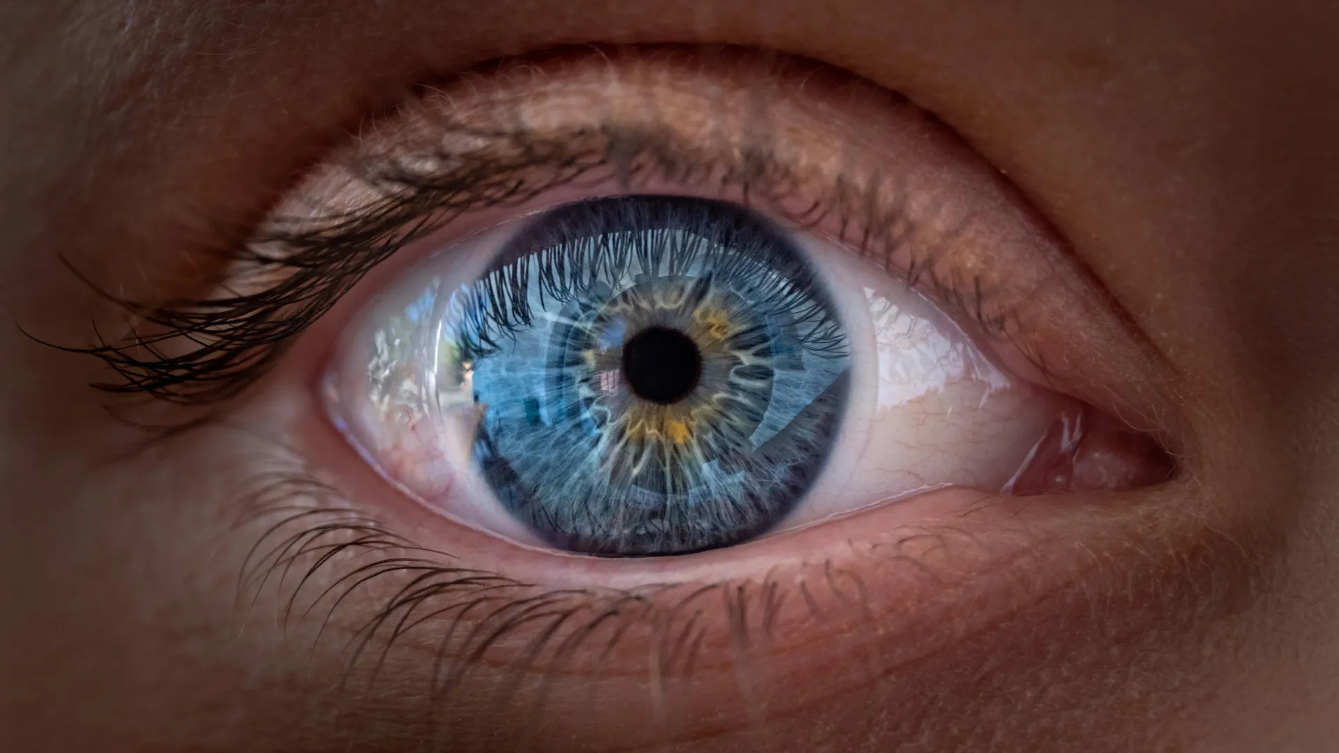
Scientists at Johns Hopkins University have made a groundbreaking discovery, revealing a surprising and previously unrecognized role for a vitamin A derivative, retinoic acid, in conjunction with thyroid hormones, in the intricate development of human vision. Their pioneering research, published in the prestigious Proceedings of the National Academy of Sciences, meticulously details how these molecules collaborate to orchestrate the formation of the foveola, the minute yet critical region of the eye responsible for our sharpest, most detailed central sight. This revelation fundamentally challenges a prevailing scientific explanation that has underpinned ophthalmological understanding for approximately 30 years, opening new avenues for both fundamental research and potential therapeutic interventions for debilitating vision loss conditions.
The core of this transformative discovery lies in an unexpected cellular metamorphosis observed within the developing human retina before birth. Researchers documented that certain specialized cells, initially programmed to detect blue light, undergo a remarkable transformation, changing their identity to become cells sensitive to red and green light. This precise and dynamic cellular reprogramming is crucial for establishing the highly specialized arrangement of photoreceptors within the foveola, which is essential for clear, high-acuity central vision—a hallmark of human sight. The implications of these findings are profound, poised to reshape scientists’ understanding of human eye development and potentially accelerate the quest for innovative treatments for conditions like macular degeneration, glaucoma, and various other disorders that severely compromise vision.
"This represents a pivotal advancement in deciphering the intricate inner workings of the center of the retina, an indispensable part of the eye and tragically, often the first to fail in individuals afflicted with macular degeneration," stated Dr. Robert J. Johnston Jr., an associate professor of biology at Johns Hopkins University and the lead author of the seminal study. "By gaining a deeper understanding of this complex region and by refining our ability to grow functional organoids that faithfully mimic its function, our long-term aspiration is to one day cultivate and transplant these specialized tissues to potentially restore lost vision."
Unlocking the Secrets of Human Vision: A Novel Approach
To systematically investigate the complex processes underlying human vision formation, Dr. Johnston and his dedicated team of colleagues employed an innovative laboratory technique they have been instrumental in developing and refining over the past several years: the cultivation of miniature human retinal tissue, aptly termed organoids. Unlike traditional research heavily reliant on animal models, which often fail to replicate the nuances of human development, these sophisticated organoids offer an unprecedented window into human biological processes.
These small, self-organizing clusters of cells, meticulously grown from fetal cells, possess the remarkable ability to reproduce key structural and functional features of developing human tissue. By observing these retinal organoids over an extended period of several months, the scientists were able to precisely track and analyze cellular changes and developmental trajectories that would otherwise be virtually impossible to study directly during the sensitive period of human pregnancy. This technological leap has proven indispensable in unraveling mysteries previously considered intractable.
The researchers’ investigative efforts were meticulously focused on the foveola, a minute depression located at the very center of the macula, within the retina. The retina itself is the light-sensitive neural tissue layer situated at the back of the eye, responsible for converting incoming light into electrical signals that the brain interprets as images. Within this critical structure, the foveola plays an exceptionally vital role in mediating detailed central vision, enabling humans to perform essential tasks such as reading fine print, recognizing familiar faces, and discerning intricate details with unparalleled clarity.
Despite its diminutive size—occupying merely a fraction of the total retinal area—the foveola is disproportionately responsible for approximately 50% of all human visual perception, underscoring its immense functional importance. Its unique cellular architecture and specialized photoreceptor distribution are what grant humans their exceptional visual acuity.
The Surprising Cellular Orchestration Behind Our Sharpest Sight
Central to comprehending the foveola’s remarkable capabilities is a specific class of specialized light-detecting cells known as cones. In contrast to rod cells, which are primarily optimized for vision in dim light and detecting motion, cones are the photoreceptors responsible for daytime vision, high-resolution perception, and, crucially, color discrimination.
Humans possess three distinct types of cone cells, each exquisitely tuned to be most sensitive to different wavelengths of light. These are conventionally categorized as blue, green, and red cones, corresponding to their peak spectral sensitivities. By intelligently comparing and integrating the signals received from these three different types of cones, the human brain can perceive and distinguish an astonishingly broad spectrum of colors, forming the rich tapestry of our visual world.
However, the distribution of these cone cells is not uniform across the entire retina. A key characteristic of the human foveola, and indeed the macula, is its unique composition: it is densely packed with red and green cones but conspicuously lacks blue cones, which are predominantly found in the periphery of the retina. This highly precise and asymmetric arrangement of cone photoreceptors is absolutely essential for the exceptionally sharp, high-resolution vision produced by the very center of the eye.
For many decades, developmental biologists and ophthalmologists have grappled with the perplexing question of how this distinctive pattern of cone distribution develops during embryogenesis. A significant impediment to answering this question has been the stark difference in retinal organization between humans and commonly studied animal models, such as mice and fish. These animals do not possess a fovea or foveola with the same specialized architecture as humans, rendering traditional animal experiments largely inadequate for investigating the developmental pathways of these specialized cells in our species.
The advent of laboratory-grown human retinas, or organoids, offered an unprecedented and powerful new opportunity to finally unravel this longstanding biological mystery, providing a direct observational platform for human-specific developmental processes.
Vitamin A and Thyroid Hormones: A Coordinated Developmental Symphony
The Johns Hopkins research team’s meticulous observations of these human retinal organoids uncovered compelling evidence that the precise arrangement of cones within the foveola develops through two exquisitely coordinated and sequential processes. Together, these processes dictate not only which types of cells initially form in the central retina but also how some of those cells undergo a remarkable change in identity as development progresses.
The chronological insights provided by the organoid model were particularly striking. During the critical period between weeks 10 and 12 of human fetal development, a small, transient population of blue cones is initially present within the nascent foveola. However, by week 14 of gestation, these blue-sensitive cells appear to have undergone a profound transformation, converting into red and green cones, thereby establishing the mature foveolar composition.
The team successfully identified two crucial chemical influences that govern and drive this intricate cellular reprogramming.
The first key player is retinoic acid, a powerful signaling molecule derived from vitamin A (retinol). Retinoic acid is a well-known master regulator of cell differentiation and specialization throughout the entire body, influencing gene expression that dictates cell fate. In the context of the developing retina, the controlled breakdown and local concentration of this molecule act to limit the initial formation of blue cones specifically within the central foveolar region. This ensures that the foveola does not become overly populated with blue cones, which would detract from its sharp vision capabilities.
The second critical influence originates from thyroid hormones. While widely recognized for their indispensable roles in regulating metabolism and overall growth, thyroid hormones also exert profound effects on cellular development and differentiation across various tissues. In this specific retinal context, these hormones appear to actively encourage and facilitate the remaining blue cones—those that did initially form in the foveola—to undergo their identity shift, transforming into red and green cones. This two-pronged attack, first limiting blue cones and then converting the existing ones, ensures the foveola achieves its optimal composition.
"It’s a beautifully orchestrated two-step process," Dr. Johnston explained. "First, retinoic acid helps establish the initial pattern by suppressing blue cone formation in the foveola. Then, thyroid hormone plays a crucial role in converting the leftover blue-sensitive cells into red and green cones. This conversion is immensely important because the presence of blue cones in that central area would significantly compromise visual acuity."
It is crucial to emphasize that this discovery pertains specifically to how vitamin A signaling influences and guides eye development before birth. The research does not imply or establish that taking vitamin A supplements can improve or enhance sharp central vision in adults, as the developmental window for this cellular transformation occurs in utero. The context of vitamin A’s role in adult vision, such as its necessity for the visual cycle in rod cells for night vision, is distinct from this newly discovered developmental function.
Challenging a 30-Year-Old Scientific Theory: A Paradigm Shift
The results of this Johns Hopkins study offer a striking and compelling alternative to a long-held scientific explanation that has significantly influenced vision research for many decades.
Under the previously dominant and longstanding model, which gained traction approximately 30 years ago, it was theorized that any blue cones that initially appeared in the developing foveola simply migrated away from this central region as the retina matured. The prevailing scientific consensus generally assumed that once these photoreceptor cells committed to becoming a particular type of cone—be it blue, green, or red—their cellular identities remained rigidly fixed and immutable throughout development.
The new findings, however, strongly suggest a fundamentally different mechanism. Rather than a simple relocation or migration, the data indicates that at least a significant proportion of these early blue cones may undergo a profound change in identity, converting into red or green cones as the eye progresses through its developmental stages. This flexibility in cell fate was largely unappreciated in the context of mature photoreceptor identity.
"The main model prevalent in the field for roughly three decades posited that the few blue cones initially found in that central region would somehow just move out of the way, based on the assumption that these cells make their definitive identity decision early on and retain that cell type forever," Dr. Johnston elaborated. "While we cannot definitively rule out some degree of cell migration, our robust data strongly supports an alternative model: that these cells actually convert their identity over time, which is truly surprising and represents a significant departure from previous thinking."
While the researchers acknowledge that some degree of cellular migration might still occur, their compelling observations provide substantial evidence for the idea that developing cone cells possess considerably more plasticity and developmental flexibility than was previously recognized by the scientific community. This newfound understanding of cellular malleability could fundamentally alter how scientists approach the study of human color vision formation and the intricate development of the retina’s most specialized and functionally critical regions.
From Discovery to Cure: The Potential for Restoring Lost Vision
Beyond its profound implications for understanding how healthy human eyes develop, this groundbreaking discovery holds immense promise for informing and accelerating the development of treatments for devastating diseases that damage the retina and cause irreversible vision loss.
One particularly promising application lies in addressing macular degeneration, a leading cause of central vision loss, especially prevalent among older adults worldwide. This progressive disease specifically damages the macula, the crucial retinal region responsible for detailed central vision, severely interfering with daily activities such as reading, driving, and recognizing faces. While certain treatments can slow or manage specific forms of the disease, there is currently no definitive cure, highlighting the urgent need for novel therapeutic strategies. Glaucoma, another major cause of blindness, involves damage to the optic nerve, often linked to high intraocular pressure, and while different in mechanism, could also benefit from advancements in understanding retinal cell health and regeneration.
A deeper understanding of the precise mechanisms by which the retina creates and organizes its specialized light-sensitive cells could eventually empower scientists to produce healthy, functional replacements in the laboratory for transplantation. This is the cornerstone of regenerative medicine.
Dr. Johnston’s team is now actively engaged in enhancing and refining their retinal organoids to more closely and accurately reproduce the complex structure and sophisticated function of the human eye. The ambitious long-term objective of this research is to develop precisely controlled populations of photoreceptors—the light-detecting cells—that are optimized for potential transplantation into damaged or diseased retinas.
Katarzyna Hussey, a key study author and former doctoral student in Dr. Johnston’s laboratory, who is now a molecular and cell biologist at CiRC Biosciences, a pioneering cell therapy company in Chicago, firmly believes that this organoid-based approach could unlock an entirely new and critical direction for vision restoration.
"The ultimate goal with leveraging this advanced organoid technology is to eventually be able to generate an almost ‘made-to-order’ population of precisely the photoreceptors needed," Hussey explained. "A major avenue of potential is cell replacement therapy, where we could introduce healthy, lab-grown cells that are capable of integrating seamlessly into the existing retinal tissue and, crucially, potentially restore that lost vision."
The core idea behind this therapeutic strategy is to introduce healthy, functionally competent cells that can successfully integrate with the eye’s existing neural circuitry, thereby replacing or augmenting functions lost due to disease or injury. Achieving this monumental goal will necessitate extensive further research and development, requiring researchers to rigorously demonstrate that transplanted cells can survive long-term, establish proper synaptic connections with the eye’s complex existing neural network, and function safely and effectively within the delicate ocular environment.
Hussey, while optimistic, judiciously emphasized that this revolutionary technology remains far from routine clinical use. She underscored that extensive development, rigorous optimization for safety and efficacy, and exhaustive testing are still absolutely necessary before such therapies could be considered for human application.
"These are inherently very long-term experiments, and naturally, we would need to conduct extensive optimizations for both safety and efficacy studies well in advance of any move into clinical trials," Hussey noted. "However, the scientific pathway laid out by this discovery represents a truly viable and promising journey toward meaningful vision restoration."
For the immediate future, this groundbreaking discovery provides something fundamentally invaluable: a significantly clearer and more detailed explanation of how the human eye develops its remarkable and unparalleled ability to perceive fine details. By revealing the intricate ballet between vitamin A signaling and thyroid hormones in shaping the foveola—the retina’s most specialized region—scientists at Johns Hopkins have taken another monumental step toward fully understanding the genesis of vision at its earliest stages and, crucially, toward developing innovative strategies to restore it when disease inevitably strikes. The journey from fundamental discovery to clinical application is long, but this research illuminates a critical path forward for millions worldwide suffering from vision impairment.
