
 By Gabriel Paijo
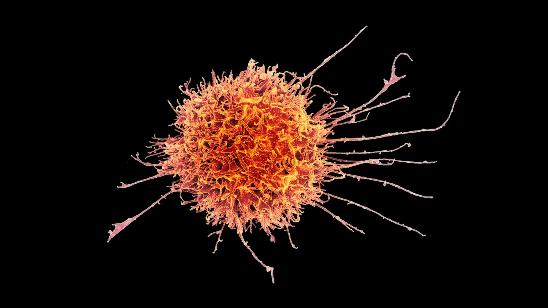
The landscape of modern oncology has been fundamentally altered by the advent of cell-based immunotherapies, particularly Chimeric Antigen Receptor (CAR) T-cell treatments. While these therapies have achieved remarkable remission rates in "liquid" cancers, such as leukemia and lymphoma, the vast majority of cancer diagnoses involve solid tumors, which have remained stubbornly resistant to similar interventions. Researchers at Stanford Medicine, in a multi-institutional collaboration, have announced a significant breakthrough in this arena by engineering a specialized form of "tissue-resident" natural killer (NK) cells. This new strategy, detailed in a study published in Science Translational Medicine, addresses the primary obstacle in treating solid tumors: the inability of immune cells to penetrate the dense, immunosuppressive environment of a tumor mass.
The Biological Fortress: Why Solid Tumors Resist Treatment
To understand the significance of the Stanford discovery, one must first recognize the physiological barriers presented by solid malignancies. Unlike blood cancers, where malignant cells circulate freely and are easily accessible to intravenous immune therapies, solid tumors—such as those found in the lungs, liver, or head and neck—function like biological fortresses. These tumors are surrounded by a complex stroma and a microenvironment that is often hypoxic and acidic, creating a physical and chemical barrier against infiltrating immune cells.
Furthermore, solid tumors frequently secrete signaling molecules, such as transforming growth factor-beta (TGF-β), which serve to "disarm" any immune cells that manage to enter. This leads to a state of exhaustion or dysfunction in the body’s natural defenses. Conventional natural killer cells, which circulate in the bloodstream, are adept at identifying and killing abnormal cells in circulation but often fail to take up permanent residence within these hostile tumor tissues.
Transforming Natural Killer Cells into Tissue-Resident Hunters
Natural killer cells were first identified in the 1970s and are categorized as part of the innate immune system. Unlike T-cells, which require a specific "primer" or exposure to an antigen to recognize a threat, NK cells are hardwired to identify and attack cells that show signs of stress or infection. This makes them an ideal candidate for universal cancer therapies.
The Stanford team, led by John Sunwoo, MD, the Edward C. and Amy H. Sewall Professor in the School of Medicine, focused on a specific subset known as tissue-resident natural killer cells. These cells are naturally found in various organs, including the skin and liver, and are uniquely adapted to survive and function within dense tissues rather than the bloodstream.
"We show that these tissue-resident natural killer cells infiltrate into the solid tumors much better than conventional natural killer cells," Sunwoo stated. "It was very reproducible, very striking and very clear."
The research team, including co-lead authors Nina Horowitz, PhD, Imran Mohammad, PhD, and June Ho Shin, PhD, sought to replicate the conditions that allow these cells to thrive within tissues. By isolating NK cells from healthy human donors and exposing them to specific stimuli, they were able to "program" the cells to adopt a tissue-resident phenotype.
The "Goldilocks" Mechanism of TGF-β Signaling
A central component of this cellular transformation involves the protein TGF-β. In many clinical contexts, TGF-β is viewed as a "bad actor" because it helps tumors grow and suppresses immune responses. However, the Stanford researchers discovered that the timing and dosage of TGF-β exposure act as a critical developmental switch for NK cells.
Sunwoo described the discovery as a "Goldilocks" scenario. If NK cells are exposed to too much TGF-β for a prolonged period, they become inhibited and dysfunctional, essentially losing their ability to kill. However, if they receive a precise, short-lived burst of the signal, they transform into highly aggressive, tissue-resident killers.
To achieve this "just right" balance, the researchers briefly exposed the circulating NK cells to short-lived human epithelial tumor cells. This direct physical contact provided the necessary TGF-β and other secondary activating signals to trigger the transformation. The resulting cells, known as cytotoxic tissue-resident NK cells, expressed specific surface proteins—CD49a, CD103, and CD39—that allow them to bind to and enter solid tumor masses.
Experimental Results and Synergistic Effects
The efficacy of these modified cells was tested in murine models bearing human-derived tumors, including melanoma and head and neck squamous cell carcinoma. The results demonstrated that the tissue-resident NK cells were significantly more effective at slowing tumor growth than their circulating counterparts.
The study also highlighted a powerful synergy between the modified NK cells and cetuximab, a monoclonal antibody currently used to treat metastatic colorectal cancer and head and neck squamous cell carcinoma. Cetuximab works by binding to the epidermal growth factor receptor (EGFR) on cancer cells, effectively "flagging" them for destruction. While cetuximab often shows limited efficacy as a monotherapy, its combination with the modified NK cells produced dramatic results.
In mice treated with the combination therapy, tumor growth was suppressed much more effectively over a 30-day period than in mice receiving either treatment alone. Observations indicated that the mice receiving the combination remained healthy and active throughout the study duration, with no apparent adverse side effects, providing a crucial "proof of concept" for future human applications.
Moving Toward an "Off-the-Shelf" Therapeutic Model
One of the most significant implications of the Stanford study is the potential for a universal, "off-the-shelf" cell therapy. Current CAR-T therapies are largely autologous, meaning they must be manufactured using a patient’s own cells. This process is prohibitively expensive, often costing hundreds of thousands of dollars per dose, and requires a weeks-long manufacturing period that some critically ill patients cannot survive.
Natural killer cells, however, are allogeneic-friendly. They do not typically cause graft-versus-host disease (GvHD), a dangerous condition where donor immune cells attack the recipient’s body. This allows for the possibility of using cells from a single healthy donor to create multiple doses for different patients.
Sunwoo and his team have developed and applied for a patent on a method to expand these modified cells at scale. They estimate that a single donor’s cells could yield approximately 20 treatment doses within two weeks. These doses can be cryopreserved and shipped to hospitals, allowing for immediate administration upon diagnosis.
"It would be almost an off-the-shelf drug," Sunwoo noted. "It could make cell therapy much more accessible to a wider variety of patients."
Chronology of Development and Future Clinical Trials
The path to this discovery has been marked by several key milestones in the Stanford lab and the broader scientific community:
- 1970s: Discovery of natural killer cells as a distinct immune population.
- 2010s: Success of CAR-T in blood cancers highlights the limitations of cell therapy for solid tumors.
- 2018-2022: The Sunwoo lab identifies the specific surface markers (CD49a, CD103) that distinguish tissue-resident NK cells.
- 2023: Researchers refine the "Goldilocks" method for TGF-β exposure using epithelial cell contact.
- Early 2024: Publication of the findings in Science Translational Medicine, detailing the efficacy of the cells in mouse models and their synergy with cetuximab.
- Late 2024 (Anticipated): Launch of a Phase I clinical trial to test the therapy in patients with advanced squamous cell carcinoma, pending FDA approval.
Broader Implications for Oncology and Immunology
The Stanford study represents a shift in focus from "circulatory immunology" to "tissue-centric immunology." For decades, blood samples have been the primary source of data for immune research because of their accessibility. However, as Sunwoo pointed out, the most critical interactions between cancer and the immune system occur within the tissues.
By mastering the transition from circulating cells to tissue-resident cells, researchers may have found a key to unlocking treatments for a wide array of cancers. If the upcoming Phase I trials prove successful, this platform could potentially be adapted for other solid tumors, such as pancreatic, lung, and breast cancers, by pairing the modified NK cells with different tumor-targeting antibodies.
The financial and logistical benefits of an off-the-shelf NK cell therapy cannot be overstated. By reducing the "vein-to-vein" time—the period between cell collection and patient infusion—this therapy could bridge the gap for patients with rapidly progressing metastatic disease. Furthermore, the ability to manufacture in batches would likely drive down the cost of immunotherapy, making it a viable option for healthcare systems globally.
While the transition from mouse models to human patients remains a significant hurdle, the Stanford team’s findings provide a robust framework for the next generation of cancer treatment. The study was supported by the National Institutes of Health, the Tai Tsun Wu Research Fund for Natural Killer Cell Immunotherapy, and the Stanford Bio-X Fellowship, with contributions from researchers at Ohio State University and Washington University School of Medicine.
