
 By Gabriel Paijo
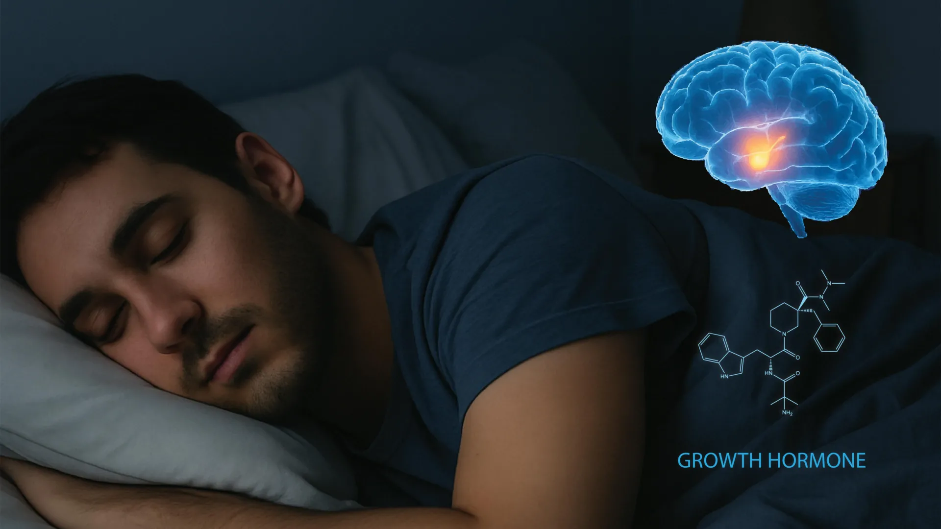
The traditional understanding of sleep as a period of physiological dormancy has been further dismantled by a landmark study from the University of California, Berkeley, which identifies the specific neural architecture responsible for the release of growth hormone during rest. Published in the prestigious journal Cell, the research identifies a sophisticated, bidirectional communication system between the brain’s sleep centers and the endocrine system. This discovery not only clarifies how deep sleep triggers the production of growth hormone (GH) but also reveals a surprising feedback loop where the hormone itself signals the brain to regulate wakefulness. By mapping these circuits, scientists have opened new avenues for treating a spectrum of conditions, ranging from childhood growth disorders and adult metabolic syndromes to neurodegenerative diseases like Alzheimer’s and Parkinson’s.
Growth hormone, a protein produced by the pituitary gland, is a master regulator of human physiology. While its role in childhood development is well-documented, its importance persists throughout the human lifespan, governing muscle protein synthesis, bone density, and the complex metabolism of fats and sugars. For decades, clinicians have observed that peak GH secretion occurs during the deepest stages of non-rapid eye movement (non-REM) sleep. However, the precise "wiring" of the brain that coordinates this surge remained one of the great mysteries of neuroendocrinology. The Berkeley team, led by Professor Yang Dan, has finally provided the blueprint for this essential biological process.
The Somatotropic Axis and the Architecture of Sleep
To understand the significance of the Berkeley study, it is necessary to examine the "somatotropic axis"—the pathway through which the brain controls growth. This axis primarily involves the hypothalamus, a small but vital region at the base of the brain, and the pituitary gland. The hypothalamus acts as a command center, releasing chemical messengers that either stimulate or inhibit the pituitary’s production of GH.
Historically, researchers relied on systemic blood sampling to track hormone levels, a method that offered a "low-resolution" view of the process. These studies confirmed that GH levels are pulsatile, with the largest pulse occurring shortly after the onset of deep sleep. Yet, because blood sampling cannot capture the millisecond-by-millisecond firing of neurons, the underlying neural triggers remained invisible. "People know that growth hormone release is tightly related to sleep, but only through drawing blood," explained Xinlu Ding, the study’s first author and a postdoctoral fellow at UC Berkeley. By using advanced electrophysiological recordings and optogenetics in mice, the team was able to observe the brain’s internal dialogue in real-time.
Identifying the Hypothalamic Gatekeepers
The research focused on a specific cluster of neurons within the hypothalamus that produce growth hormone-releasing hormone (GHRH). These neurons are the "gas pedal" for GH production. Conversely, the team examined neurons that produce somatostatin, which acts as the "brake."
The Berkeley study revealed that these two cell populations operate with remarkable coordination during different sleep stages. During non-REM sleep—the restorative phase characterized by slow brain waves—somatostatin activity drops significantly, removing the inhibition on the pituitary gland. Simultaneously, GHRH neurons increase their activity, signaling the pituitary to flood the bloodstream with growth hormone.
The researchers also observed a curious phenomenon during REM sleep, the stage associated with vivid dreaming. In this stage, both GHRH and somatostatin neurons increase their activity. While these hormones have opposing functions, their synchronized firing during REM appears to facilitate a specific pattern of GH release that differs from the steady surge seen in non-REM sleep. This suggests that every stage of sleep plays a distinct, calculated role in maintaining hormonal balance.
The Discovery of a Bidirectional Feedback Loop
The most groundbreaking aspect of the study is the discovery that growth hormone does not merely respond to sleep; it actively influences it. The researchers identified a feedback loop involving the locus coeruleus (LC), a nucleus in the brainstem that serves as the primary source of norepinephrine, the chemical responsible for arousal, alertness, and the "fight or flight" response.
As growth hormone levels rise during the night, the hormone travels back to the brain and binds to receptors on the neurons of the locus coeruleus. This interaction gradually increases the excitability of the LC. When GH levels reach a certain threshold, the LC’s activity helps transition the brain from sleep to wakefulness.
"This suggests that sleep and growth hormone form a tightly balanced system," noted Daniel Silverman, a co-author of the study. "Too little sleep reduces growth hormone release, and too much growth hormone can in turn push the brain toward wakefulness." This homeostatic mechanism ensures that the body receives enough GH for repair and growth while also ensuring the individual eventually wakes up to face the day’s demands.
Experimental Methodology: Optogenetics and Murine Models
The study utilized mouse models, which are indispensable in sleep research due to their highly conserved hypothalamic functions. While humans are monophasic sleepers (sleeping in one long block), mice are polyphasic, sleeping in short bursts throughout a 24-hour cycle. This allowed the researchers to observe the transition between sleep and wakefulness hundreds of times, providing a robust dataset that would be impossible to achieve in human subjects.
Using optogenetics—a technique that involves using light to turn specific neurons on or off—the team was able to prove causality. When they artificially stimulated GHRH neurons, they observed an immediate spike in GH levels. When they blocked the feedback to the locus coeruleus, the mice experienced disrupted sleep-wake transitions, proving that the GH-to-LC pathway is essential for normal sleep architecture.
Clinical Implications: From Metabolic Disease to Neurodegeneration
The implications of mapping this circuit extend far beyond basic biology. Disrupted sleep is a hallmark of "Metabolic Syndrome," a cluster of conditions including obesity, high blood pressure, and high blood sugar. Since GH is a primary regulator of fat metabolism and glucose levels, chronic sleep deprivation creates a vicious cycle. Lower GH levels lead to increased fat storage and insulin resistance, which in turn can lead to sleep apnea and further sleep disruption.
Furthermore, the link to the locus coeruleus is particularly significant for neurology. The LC is one of the first areas of the brain to show pathology in Alzheimer’s and Parkinson’s diseases. Degeneration of the LC leads to the profound sleep disturbances and "sundowning" often seen in these patients. By understanding how growth hormone interacts with the LC, scientists may be able to develop hormonal therapies that stabilize this region of the brain, potentially slowing cognitive decline or improving the quality of life for patients with neurodegenerative disorders.
"Growth hormone not only helps you build your muscle and bones… but may also have cognitive benefits, promoting your overall arousal level when you wake up," Ding stated. This suggests that the "grogginess" associated with poor sleep may be partially due to an interrupted GH feedback loop, failing to properly "prime" the locus coeruleus for morning alertness.
The Evolution of Sleep Science
This study marks a significant milestone in a timeline of discovery that began in the late 1960s. In 1968, researchers first noted the correlation between slow-wave sleep and GH secretion. For the next fifty years, the scientific community understood the what and the when, but the how remained elusive.
The Berkeley research contextualizes sleep as an active metabolic state. It challenges the "waste product" theory of sleep—the idea that sleep is merely for clearing out neural debris—and positions it as a period of active endocrine management. The discovery of the GH-LC feedback loop adds a new layer to our understanding of homeostasis, the body’s ability to maintain a stable internal environment.
A New Frontier in Hormonal and Sleep Therapy
The researchers are optimistic that their findings will lead to "precision" treatments. Current growth hormone therapies are often systemic and can have significant side effects. By targeting the specific hypothalamic neurons or the LC receptors identified in this study, future gene therapies or pharmacological interventions could restore hormonal balance without affecting other systems.
"This circuit could be a novel handle to try to dial back the excitability of the locus coeruleus," Silverman suggested, referring to potential treatments for insomnia or hyper-arousal disorders. Conversely, for children with growth deficiencies, ensuring the integrity of this neural circuit might be as important as the hormone levels themselves.
While the study was conducted in mice, the fundamental structures involved—the hypothalamus, the pituitary, and the locus coeruleus—are remarkably similar across all mammals. The researchers anticipate that future human trials, perhaps utilizing high-resolution fMRI and advanced endocrine monitoring, will confirm these pathways in the human brain.
In conclusion, the UC Berkeley study transforms our understanding of the relationship between the pillow and the pituitary. It reaffirms the ancient wisdom that sleep is the "great restorer," but adds a modern caveat: sleep is also a great regulator. As society continues to grapple with a "sleep loss epidemic," the mapping of this neural circuit serves as a potent reminder that every hour of lost sleep is not just a loss of rest, but a disruption of the very chemical messengers that build our bodies and sharpen our minds.
