
 By Lina carolina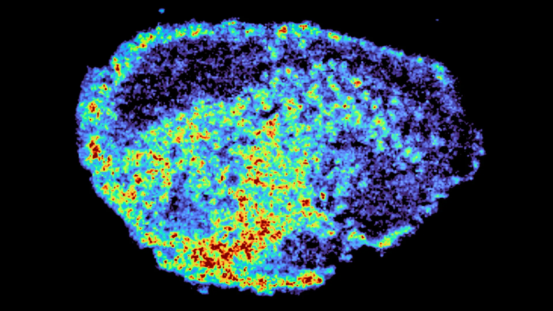
A groundbreaking study published in the prestigious journal Nature Communications has illuminated a critical, previously underappreciated factor dictating the success of targeted cancer therapies. Researchers at the MRC Laboratory of Medical Sciences (LMS), led by Dr. Louise Fets, have pinpointed the accumulation of PARP inhibitors within cellular lysosomes as a significant driver of treatment variability, explaining why these potent drugs can be a lifeline for some patients while proving ineffective for others. This discovery holds profound implications for the future of personalized cancer medicine, potentially paving the way for more precise and effective therapeutic strategies.
The challenge of differential treatment response has long been a formidable hurdle in oncology. While advancements in precision medicine have yielded remarkable progress, the inherent biological complexity of tumors means that even highly targeted therapies can exhibit a spectrum of efficacy across individuals. This variability is particularly pronounced with PARP inhibitors, a class of drugs that have revolutionized the treatment landscape for ovarian, breast, and prostate cancers by exploiting specific DNA repair pathway deficiencies in cancer cells. The fundamental principle behind their action is to induce lethal DNA damage by inhibiting Poly(ADP-ribose) polymerase (PARP) enzymes, which are crucial for repairing single-strand DNA breaks. When PARP is inhibited, these breaks accumulate, leading to double-strand breaks during DNA replication that cancer cells, especially those with compromised homologous recombination repair (HRR) pathways, struggle to fix, ultimately triggering programmed cell death (apoptosis). However, the clinical reality often falls short of this ideal, with a significant proportion of patients either not responding to treatment or developing resistance over time. Understanding the intricate journey of these drugs within the tumor microenvironment has been a persistent enigma, hindering efforts to optimize their use.
Mapping the Unseen Journey: Advanced Imaging Unveils Drug Distribution
The LMS research team employed cutting-edge imaging technologies to meticulously track the distribution of PARP inhibitors within patient-derived ovarian tumor samples. This novel approach allowed them to visualize, at an unprecedented level of detail, how these drugs navigate the complex cellular architecture of a tumor. By utilizing thin slices of ovarian tumors, known as "explants," maintained in a viable state ex vivo, the researchers could directly observe drug uptake and distribution in real human tumor tissue under controlled laboratory conditions. This method offers a significant advantage over traditional cell culture models, which may not fully recapitulate the intricate three-dimensional structure and cellular heterogeneity of native tumors.
A cornerstone of their methodology was mass spectrometry imaging (MSI). This powerful technique enabled the precise spatial mapping of drug molecules within the tumor tissue, revealing areas of high and low drug concentration with remarkable accuracy. MSI generates detailed chemical maps, essentially painting a picture of where specific molecules are located within a sample. This was powerfully complemented by spatial transcriptomics, a technology that allows for the simultaneous analysis of gene expression patterns within the same tissue slice, correlating cellular activity with drug localization. By overlaying these datasets, the scientists could identify specific regions within the tumor that had accumulated high drug levels and simultaneously examine the genetic signatures of the cells in those areas, as well as in adjacent regions with lower drug concentrations.
The findings were striking. The researchers observed significant disparities in drug distribution, not only between different tumor samples from distinct patients but also within individual tumors. Even when subjected to the same drug dosage, the concentration of PARP inhibitors varied considerably from cell to cell and from region to region within the tumor mass. This heterogeneity in drug exposure was a pivotal revelation, suggesting that differences in how drugs penetrate and disseminate within the tumor microenvironment could be a major determinant of treatment outcome.
"A novel aspect of this study was the use of mass spectrometry imaging to directly measure and visualize drug uptake in patient tumour tissue," stated Dr. Zoe Hall, a senior author on the study and Associate Professor at Imperial’s Department of Metabolism, Digestion and Reproduction. "Through the spatial mapping of drug molecules, we could pinpoint regions of high and low drug and compare gene expression, from the same tissue slice, using spatial transcriptomics." This integrated approach provided a comprehensive view of drug dynamics and cellular response, moving beyond simple measurements of drug concentration to understanding the functional implications of that distribution.
Lysosomes: The Unexpected Drug Reservoirs
The investigation into the mechanisms driving this uneven drug distribution led to a remarkable discovery: the central role of lysosomes. These small, membrane-bound organelles within cells are often described as the cell’s "recycling centers," responsible for degrading waste materials and cellular debris. However, the LMS study revealed that lysosomes can also act as unintended, dynamic reservoirs for certain PARP inhibitors.
The researchers found that some PARP inhibitors are actively transported into lysosomes and become sequestered within these compartments. Instead of freely circulating throughout the cell to reach their targets, these drugs accumulate inside lysosomes, creating localized "hotspots" of high drug concentration. This lysosomal sequestration effectively transforms these organelles into slow-release depots. While this can lead to prolonged drug exposure in certain cellular environments, it also means that other areas of the tumor, or even adjacent cells within the same tissue, may receive significantly lower drug doses.
This mechanism of lysosomal storage is not universal across all PARP inhibitors. The study specifically identified that drugs like rucaparib and niraparib are susceptible to this lysosomal accumulation. In contrast, olaparib, another widely used PARP inhibitor, did not exhibit the same degree of sequestration within lysosomes. This differential behavior suggests that the chemical properties of specific PARP inhibitors play a crucial role in their interaction with cellular organelles.
"We were surprised to see large variability in drug accumulation at the single-cell level," commented Dr. Carmen Ramirez Moncayo, the study’s first author and a Postdoctoral Researcher at the LMS. "This variability was driven by the build-up of a drug in lysosomes, which are acting as reservoirs, increasing the exposure of cancer cells to drugs, by storing and releasing the drug when needed." This finding offers a tangible explanation for the observed discrepancies in drug efficacy, linking cellular organelle function directly to therapeutic outcomes.
Timeline of Discovery and Methodological Evolution
The journey to this significant discovery can be traced back to the growing recognition of PARP inhibitors’ transformative potential in gynecological oncology. Following their initial success in ovarian cancer, the need to understand response variability became increasingly urgent. The development of advanced imaging techniques, such as mass spectrometry imaging and spatial transcriptomics, in recent years provided the technological impetus for this in-depth investigation.
The research likely began with the collection of patient tumor samples, a critical step that ensures the study’s relevance to real-world clinical scenarios. These samples would have undergone rigorous processing to be maintained as viable explants. The subsequent phases would have involved the controlled administration of various PARP inhibitors to these explants, followed by the application of MSI to map drug distribution. Simultaneously, spatial transcriptomics would have been employed to analyze gene expression in relation to these drug maps. The integration and analysis of these complex datasets would have then led to the identification of lysosomal sequestration as a key mechanism. The comparison of different PARP inhibitors would have been an iterative process, building upon initial observations to identify drug-specific behaviors.
Broader Impact and Future Directions: Towards Precision Oncology
The implications of this research extend far beyond ovarian cancer. PARP inhibitors are increasingly being explored and utilized in a range of other malignancies, including breast, prostate, pancreatic, and certain types of lung cancer. A deeper understanding of how these drugs are internalized, distributed, and potentially sequestered within tumor cells opens up new avenues for optimizing their clinical application.
This study suggests that factors influencing lysosomal function and drug-lysosome interactions could become critical biomarkers for predicting treatment response. If lysosomal sequestration is a significant determinant of efficacy, then strategies to modulate lysosomal activity or to design PARP inhibitors less prone to lysosomal trapping could lead to enhanced treatment outcomes.
"By understanding how drugs are taken up into cells, we can understand whether this influences why cancer drugs work for some people and not for others," explained Dr. Louise Fets, a senior author and Head of the LMS’ Drug Transport and Tumour Metabolism Group. "Eventually, we hope to be able study the molecular signature of a patient’s tumor to help to tailor therapeutic approaches in a more personalized way." This vision aligns perfectly with the overarching goals of precision oncology, which aims to tailor medical treatment to the individual characteristics of each patient.
The current study, while groundbreaking, was conducted using tumor tissue maintained outside the body. In a living patient, drug delivery is primarily via the bloodstream, and tumor vasculature is often abnormal and disorganized, further complicating drug penetration and distribution. These vascular abnormalities can create hypoxic regions within tumors, potentially influencing both drug delivery and cellular metabolism, including lysosomal function. Therefore, future research will be crucial in translating these findings from the laboratory to the clinic. Studies involving animal models and larger patient cohorts are planned to investigate how drug delivery dynamics, tumor structural complexity, and lysosomal storage mechanisms interact in the clinical setting. This will include exploring these phenomena in the context of relapsed cancers, where treatment resistance is often a significant challenge.
Supporting Data and Context
The development and approval of PARP inhibitors have been a landmark achievement in cancer therapy. For instance, olaparib was first approved by the U.S. Food and Drug Administration (FDA) in 2014 for certain types of ovarian cancer, and its indications have since expanded significantly. Rucaparib and niraparib followed, offering further options for patients. The global market for PARP inhibitors is substantial and projected to grow, underscoring their clinical importance. Despite their efficacy, response rates in unselected populations can vary. For example, in some platinum-sensitive ovarian cancer settings, response rates can range from 30% to 70%, with significant individual variation. Understanding the drivers of this variability is therefore of paramount clinical and economic importance.
The research was supported by substantial funding from key medical research bodies, including the Medical Research Council, Cancer Research UK, the Integrative Toxicology Training Partnership administered by the MRC Toxicology Unit, and a Victoria’s Secret Global Fund for Women’s Cancers Career Development Award, in partnership with Pelotonia and AACR. This multidisciplinary funding demonstrates the broad recognition of the importance of this research area.
Official Responses and Expert Commentary (Inferred)
While direct quotes from external parties were not provided in the original text, the publication of this research in Nature Communications suggests it has undergone rigorous peer review and is likely to be met with significant interest from the oncology community. Leading oncologists and researchers in the field of DNA repair and targeted therapy are expected to acknowledge the study’s contribution to understanding drug disposition. Discussions are likely to emerge regarding the potential for diagnostic tools to assess lysosomal activity in tumors or to stratify patients based on their likelihood of benefiting from PARP inhibitors that are prone to lysosomal sequestration. Pharmaceutical companies developing PARP inhibitors may also leverage these findings to inform the design of next-generation drugs with improved pharmacokinetic profiles.
In conclusion, the LMS study represents a significant leap forward in unraveling the complex interplay between drug behavior and cellular machinery in cancer treatment. By identifying lysosomes as key players in the variable efficacy of PARP inhibitors, researchers have opened a critical window into the personalized treatment of cancer. The future holds the promise of translating these intricate cellular mechanisms into tangible clinical benefits, offering hope for more effective and tailored cancer therapies for patients worldwide.
