
 By Nana O
Researchers at Weill Cornell Medicine and the Hospital for Special Surgery (HSS) have achieved a significant breakthrough in regenerative medicine, identifying a previously elusive population of stem cells that serve as the primary source for the body’s tendons and ligaments. These crucial connective tissues, responsible for linking muscles to bones, have long presented a challenge for scientific understanding due to their complex cellular makeup. This groundbreaking discovery not only sheds light on the fundamental mechanisms of musculoskeletal development and repair but also opens promising new avenues for treating debilitating conditions like lumbar spinal stenosis.
The study, published on September 7th in the prestigious journal Cell, marks a pivotal moment in the field, potentially revolutionizing how we approach connective tissue disorders. For decades, scientists have sought to pinpoint the specific progenitor cells that give rise to the diverse array of cells found in tendons and ligaments. While various candidate cell types have been proposed, none had definitively demonstrated the capacity for both self-renewal and the generation of the full spectrum of mature tendon and ligament cells. This new research fills that critical gap, identifying a single, highly specialized stem cell population with these essential characteristics.
A Deep Dive into Connective Tissue Genesis
The journey to this discovery began with the meticulous work of a research team led by co-corresponding authors Dr. Matthew Greenblatt, a pathology and laboratory medicine associate professor at Weill Cornell and pathologist at NewYork-Presbyterian/Weill Cornell Medical Center, and Dr. Sravisht Iyer, an associate professor of orthopedics at Weill Cornell and a spine surgeon at HSS. Dr. Greenblatt’s lab had a history of success in identifying key skeletal stem cell populations. Previously, his team had pinpointed the stem cell responsible for fracture repair in the outer layer of bone and later identified cells involved in the formation of the skull and spine. However, unraveling the origins of tendons and ligaments proved to be a considerably more intricate endeavor.
"Unlike bone, these tissues contain many fibroblast-like cells that look similar, making it challenging to distinguish one cell population from another," explained Dr. Greenblatt. The researchers employed advanced single-cell sequencing technologies to analyze thousands of individual cells, meticulously sorting them into distinct cell types. Through this process, they were able to identify a rare population exhibiting the defining hallmarks of "stemness"—the ability to indefinitely replicate itself while also differentiating into the various mature cell types required to build and maintain tendon and ligament structures.
In initial studies using mouse models, these identified stem cells were found residing in a specific niche within tendons and ligaments, acting as a reservoir crucial for tissue growth and repair. This observation provided a critical foundation for the subsequent investigation in human tissues.
From Bench to Bedside: Translating Discovery to Human Health
The research team then leveraged their findings from mice to meticulously search for analogous cells in human connective tissues. Human ligament samples were obtained from tissue removed during surgical procedures performed by Dr. Iyer, with full informed consent from patients. This direct access to human tissue was instrumental in validating the initial discoveries.
"We analyzed thousands of individual cells and sorted them into individual cell types. Then we identified which one had the properties we associate with ‘stemness’," Dr. Greenblatt elaborated. The researchers, including first author Dr. Lingling Hu, a postdoctoral fellow in both Dr. Greenblatt’s and Dr. Iyer’s labs, successfully confirmed that these human cells possessed the dual capacity for self-renewal and the generation of ligament cells.
A significant finding was the widespread distribution of these stem cells. "We looked in the kneecap ligament; we looked at the Achilles tendon; and everywhere we looked, we found this cell," stated Dr. Greenblatt. "So, we think this is the universal stem cell for tendons and ligaments throughout the body." This universal nature suggests a fundamental role for these cells in the development and maintenance of all fibrous connective tissues.
Lumbar Spinal Stenosis: A New Mechanistic Understanding
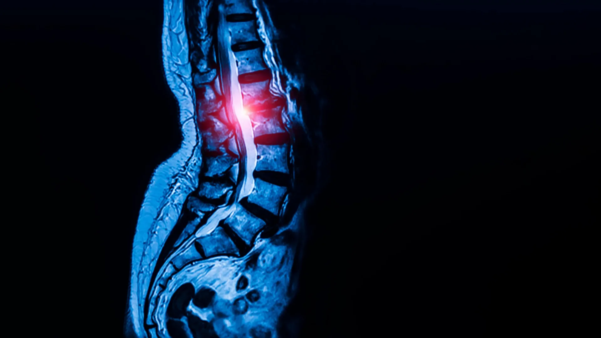
Beyond their role in normal tissue development, the researchers delved into the potential involvement of these newly identified stem cells in pathological conditions, with a particular focus on lumbar spinal stenosis. This prevalent condition, affecting an estimated 103 million people worldwide, occurs when the ligaments in the lower spine thicken, narrowing the spinal canal and potentially compressing vital nerves. The resulting symptoms can include chronic pain, numbness, and significant mobility challenges, often leaving patients with limited treatment options beyond invasive surgery once the condition becomes severe.
To investigate this link, the team compared stem cells isolated from the spinal ligaments of patients diagnosed with lumbar spinal stenosis to those collected from individuals with herniated discs but no evidence of stenosis. Pre-operative informed consent was obtained from all participants. The analysis revealed a marked increase in the abundance of the newly identified stem cells within the ligaments of individuals with spinal stenosis.
Further experiments involved transplanting these stem cells into mouse models. Stem cells derived from stenotic human ligaments consistently produced more tendon cells compared to stem cells from healthy ligaments. "Though spinal stenosis is a complex condition, this really showed us that these cells are contributing to the pathology," Dr. Greenblatt affirmed. This finding provides compelling evidence that an overactivity or dysregulation of these specific stem cells may be a key driver of ligamentous thickening in lumbar spinal stenosis.
Unraveling the Molecular Underpinnings: Calcium Signaling Emerges as a Key Player
The research team then focused on understanding the molecular mechanisms underlying this increased stem cell activity in spinal stenosis. They observed that stem cells associated with the condition exhibited heightened calcium signaling compared to their healthy counterparts. Calcium signaling is a fundamental cellular communication pathway that regulates a myriad of cellular processes, including growth and activity.
Intriguingly, by genetically enhancing calcium signaling in stem cells derived from healthy ligaments, the researchers were able to induce excessive tissue growth, mimicking the pathological changes seen in spinal stenosis. Conversely, when they reduced calcium signaling in a mouse model of lumbar spinal stenosis, the abnormal cell growth was significantly attenuated.
This bidirectional manipulation of calcium signaling underscores its critical role in regulating the behavior of these tendon and ligament stem cells and their contribution to spinal stenosis. The findings suggest that aberrant calcium signaling could be a central mechanism driving the excessive ligament thickening characteristic of the condition.
A Promising Therapeutic Horizon: Targeting Calcium Channels
The discovery that calcium signaling is a key driver of pathological ligament growth in spinal stenosis opens up a potentially revolutionary therapeutic avenue. The research points towards the possibility of targeting calcium signaling pathways to manage or even reverse the progression of the disease.
"That finding suggests calcium signaling could become a therapeutic target," stated Dr. Iyer. "It also raises the possibility that calcium channel blockers, a class of drugs already widely used to treat high blood pressure, could one day be studied as a treatment for spinal stenosis."
Calcium channel blockers are a well-established class of medications that work by inhibiting the influx of calcium ions into cells, thereby reducing cellular excitability and muscle contraction. Their existing safety profile and widespread availability make them an attractive candidate for repurposing in the treatment of spinal stenosis. However, the researchers emphasize that extensive clinical trials will be necessary to determine the safety and efficacy of such an approach in human patients.
"This is probably the first work that’s shown a potential therapeutic target for one of the most common spine conditions in the world," Dr. Iyer remarked, highlighting the profound implications of this research for millions of individuals suffering from debilitating back pain and mobility issues.
Broader Implications: Beyond Spinal Stenosis
The impact of this discovery is poised to extend far beyond lumbar spinal stenosis, offering new insights and potential therapeutic strategies for a range of other connective tissue disorders. Dr. Greenblatt indicated plans to investigate whether these same stem cells are implicated in conditions such as Marfan syndrome, a genetic disorder that affects connective tissues throughout the body, leading to a variety of serious health complications.
Furthermore, the identification of the universal stem cell for tendons and ligaments may help researchers unravel the persistent mystery behind why certain tendon and ligament injuries are notoriously difficult to heal. Conditions like rotator cuff tears, Achilles tendon injuries, and chronic tendon degeneration, which affect athletes and non-athletes alike, could potentially be addressed by therapies aimed at modulating the behavior of these foundational stem cells.
"Given that this cell appears to be the ultimate origin of all tendon and ligament cells, defects in this cell are likely at the heart of a wide range of tendon and ligament disorders," Dr. Greenblatt concluded. This broader perspective suggests that this fundamental discovery could pave the way for advancements in treating a spectrum of musculoskeletal ailments, improving healing outcomes and enhancing the quality of life for countless individuals.
Future Directions and Research Support
The research was made possible through substantial funding from a variety of sources, including the Marfan Foundation, the Children’s Tumor Foundation, the Arthritis National Research Foundation, the National Institutes of Health, and the National Research Foundation of Korea, among others. This diverse support underscores the broad scientific and medical interest in this transformative work.
The next steps will involve rigorous preclinical testing of calcium channel blockers in animal models of spinal stenosis and other connective tissue disorders. Following successful preclinical studies, human clinical trials will be initiated to evaluate the therapeutic potential of these drugs. Researchers are also keen to explore other therapeutic strategies that might target calcium signaling pathways or directly modulate the activity of these newly identified stem cells.
The identification of this pivotal stem cell population represents a significant leap forward in our understanding of connective tissue biology and disease. It offers a tangible hope for developing more effective, less invasive treatments for conditions that currently pose substantial challenges to patients and healthcare providers alike. The collaborative efforts of Weill Cornell Medicine and the Hospital for Special Surgery have not only advanced fundamental scientific knowledge but have also laid the groundwork for a new era of regenerative therapies for a wide range of musculoskeletal conditions.
