
 By Nana O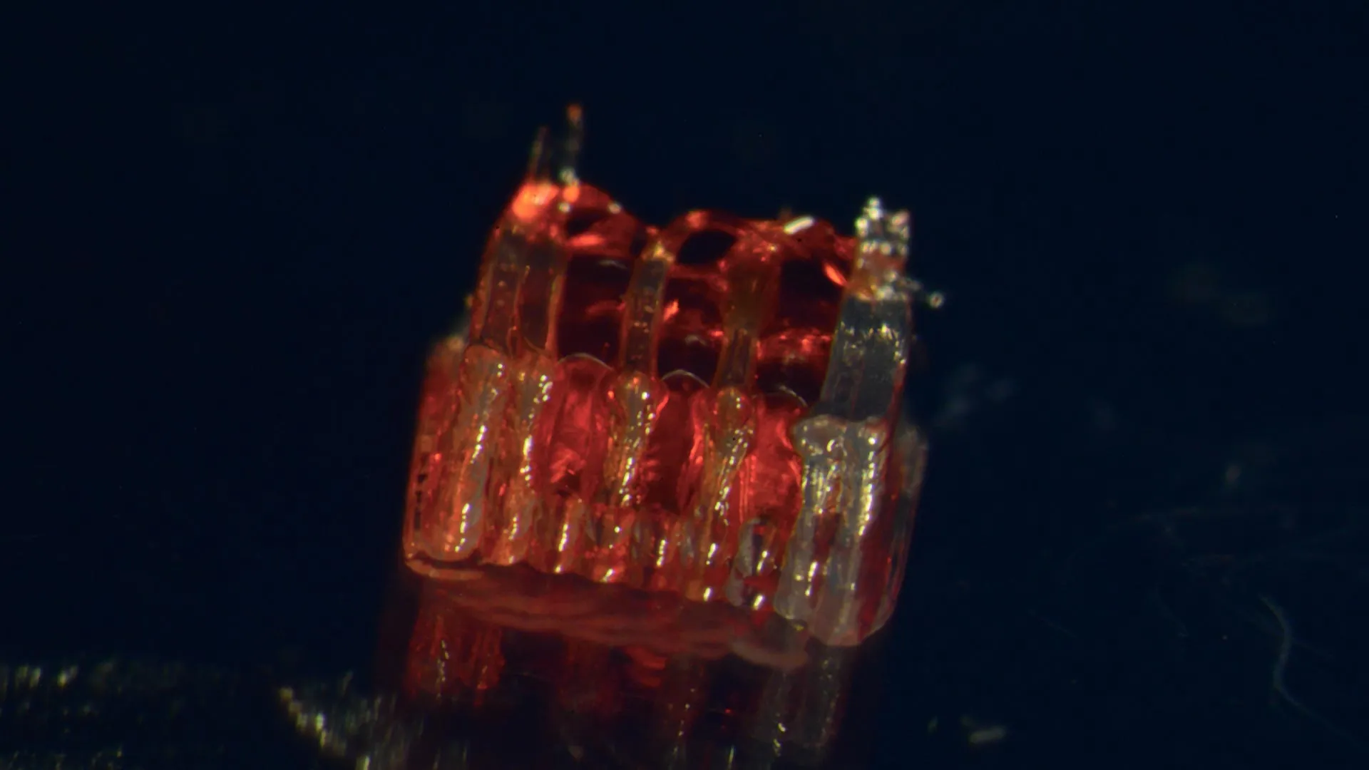
A groundbreaking advancement in regenerative medicine, developed by a research team at the University of Minnesota Twin Cities, promises a new era of hope for individuals suffering from spinal cord injuries. For the first time, scientists have successfully combined three cutting-edge technologies – 3D printing, stem cell biology, and the creation of lab-grown tissues – to demonstrate a viable pathway toward repairing severed spinal cords and restoring lost function. This pioneering work, detailed in the latest issue of the peer-reviewed scientific journal Advanced Healthcare Materials, addresses one of the most devastating and persistent challenges in modern medicine.
The Scale of the Spinal Cord Injury Crisis
Spinal cord injuries (SCIs) represent a profound public health concern, leaving hundreds of thousands of individuals in the United States facing life-altering paralysis and loss of sensation. According to the National Spinal Cord Injury Statistical Center, more than 300,000 people in the U.S. currently live with the effects of these injuries. The stark reality is that current medical interventions offer no complete reversal of the damage, and the inherent inability of damaged nerve cells to regenerate across the injury site has long been a formidable barrier to recovery. This fundamental limitation stems from the death of critical nerve cells and the failure of nerve fibers, known as axons, to regrow and bridge the gap created by the injury. It is this complex biological puzzle that the University of Minnesota team has directly confronted with their innovative approach.
A Novel Bio-Integrated Scaffold for Neural Regeneration
The core of this revolutionary technique lies in the creation of a sophisticated, bio-integrated scaffold. Utilizing advanced 3D printing technology, researchers fabricated a custom framework designed to mimic the intricate architecture of the spinal cord. This "organoid scaffold" is not merely a passive support structure; it features a network of microscopic channels, meticulously engineered to guide the growth of specialized cells.
The next critical step involves populating these channels with regionally specific spinal neural progenitor cells (sNPCs). These are not just any cells; they are derived from human adult stem cells, possessing the remarkable potential to divide and differentiate into the precise types of mature cells needed for spinal cord function, including neurons. This targeted cell sourcing is crucial for ensuring the regenerated tissue possesses the correct cellular composition and connectivity.
Directing Neural Growth: The Power of 3D Printing
The strategic integration of 3D printing with stem cell biology is what sets this research apart. "We use the 3D printed channels of the scaffold to direct the growth of the stem cells, which ensures the new nerve fibers grow in the desired way," explained Guebum Han, a former University of Minnesota mechanical engineering postdoctoral researcher and the first author on the paper, who is now with Intel Corporation. "This method creates a relay system that when placed in the spinal cord bypasses the damaged area."
This directed growth is paramount. In a healthy spinal cord, nerve fibers extend in precise directions to form complex neural circuits. When these circuits are disrupted by injury, simply implanting cells is insufficient. The 3D-printed channels act as a biological roadmap, physically guiding the regenerating axons along predetermined pathways, mimicking the natural development of the spinal cord. This precise guidance is believed to be a key factor in overcoming the chaotic and often unsuccessful attempts at regeneration observed in previous research.
Pre-Clinical Success: Promising Results in Animal Models
To rigorously test their innovative approach, the research team conducted a critical series of experiments. They transplanted these meticulously engineered scaffolds, populated with sNPCs, into rats whose spinal cords had been completely severed. The results observed were nothing short of remarkable.
Within the transplanted scaffolds, the sNPCs successfully differentiated into mature neurons. More importantly, these newly formed nerve fibers exhibited an unprecedented ability to extend in both directions – rostral (towards the head) and caudal (towards the tail) – from the implant site. This bidirectional growth is essential for re-establishing connections with the host’s existing nerve circuits, effectively bridging the severed gap.
Over time, a crucial aspect of the study involved observing the integration of these new nerve cells. The research demonstrated that the regenerated nerve cells seamlessly integrated into the host spinal cord tissue. This integration was not superficial; it led to significant functional recovery in the rats. While the specifics of the functional recovery are detailed in the full study, the implication is that the rats regained some degree of motor control or sensory perception that was previously lost due to the complete severance of their spinal cords. This preclinical success provides strong evidence for the efficacy of the combined 3D printing and stem cell approach.
A New Dawn for Regenerative Medicine
The implications of this research extend far beyond the laboratory setting. Ann Parr, a professor of neurosurgery at the University of Minnesota and a senior researcher on the project, expressed her enthusiasm for the future potential of this work. "Regenerative medicine has brought about a new era in spinal cord injury research," she stated. "Our laboratory is excited to explore the future potential of our ‘mini spinal cords’ for clinical translation."
The term "mini spinal cords" aptly describes the lab-grown organoid structures. The ability to create functional, self-organizing neural tissues in vitro that can then be integrated into a damaged nervous system represents a significant leap forward. This research moves the field closer to the long-sought goal of truly regenerative therapies for spinal cord injuries, shifting the paradigm from managing symptoms to actively repairing the underlying damage.
Path Forward: Challenges and Future Directions
While this study marks a monumental achievement, the researchers are clear that this is the beginning of a long journey toward clinical application. The current research is in its nascent stages, and significant hurdles remain before human trials can be contemplated. Scaling up the production of these complex organoid scaffolds and ensuring their long-term safety and efficacy in larger animal models and eventually humans will require extensive further research and development.
The team’s immediate goals involve optimizing the scaffold design, refining the differentiation protocols for sNPCs, and conducting more comprehensive pre-clinical studies to assess the durability and extent of functional recovery. The ultimate vision is to develop a robust and reproducible treatment that can be widely accessible to individuals affected by spinal cord injuries. This combination of advanced manufacturing and cutting-edge biological science holds immense promise for a future where paralysis due to spinal cord injury is no longer a permanent sentence.
A Collaborative Effort with Broad Support
This groundbreaking research is the product of a multidisciplinary collaboration involving experts from various departments at the University of Minnesota and beyond. Key contributors to this study include:
- From the University of Minnesota Department of Mechanical Engineering: Guebum Han and Michael McAlpine.
- From the University of Minnesota Department of Neurosurgery: Ann Parr, Hyunjun Kim, Nicolas S. Lavoie, Nandadevi Patil, and Olivia G. Korenfeld.
- From the University of Minnesota Department of Neuroscience: Manuel Esguerra.
- From Virginia Commonwealth University, Department of Physics: Daeha Joung.
The significant undertaking was made possible through substantial funding from prestigious organizations dedicated to advancing medical research. This includes grants from the National Institutes of Health (NIH), the State of Minnesota Spinal Cord Injury and Traumatic Brain Injury Research Grant Program, and the Spinal Cord Society. Such robust support underscores the national and international recognition of the importance and potential impact of this research.
The full details of this transformative study can be found in the Advanced Healthcare Materials journal, under the title, "3D-Printed Scaffolds Promote Enhanced Spinal Organoid Formation for Use in Spinal Cord Injury." This publication represents a critical milestone, making the detailed findings available to the global scientific community for further scrutiny, replication, and advancement. The work offers a beacon of hope, illuminating a path toward a future where the devastating consequences of spinal cord injuries can be effectively treated and potentially reversed.
