
 By Budi Oetomo Narendra
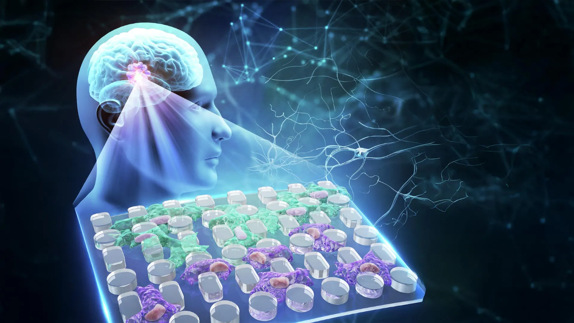
Glioblastoma, one of the most aggressive and lethal forms of brain cancer, presents an extraordinary challenge to medical science. Its notoriously poor prognosis, with a median survival often measured in mere months, stems largely from its highly infiltrative nature, making complete surgical removal virtually impossible and recurrence almost inevitable. In a surprising turn that redefines our understanding of cancer invasion, scientists have uncovered a novel mechanism by which glioblastoma cells not only navigate but actively exploit the physical properties of their surroundings to enhance their invasiveness. Contrary to expectations that a denser environment would impede cellular movement, researchers have found that the unusually thick fluid at the outer edges of glioblastoma tumors forces cancer cells to adapt, transforming them into smaller, more flexible, and ultimately more aggressive entities. This discovery, facilitated by an innovative microfluidic chip, suggests that fluid viscosity is not merely a passive barrier but an active influencer of cancer cell behavior, potentially opening new avenues for therapeutic intervention.
Glioblastoma: A Formidable Adversary in Oncology
Glioblastoma multiforme (GBM) stands as the most common and deadliest primary malignant brain tumor in adults. Characterized by rapid growth, widespread infiltration into healthy brain tissue, and resistance to conventional therapies, GBM typically carries a median survival rate of approximately 15 to 18 months, even with aggressive treatment protocols involving surgery, radiation, and chemotherapy. The tumor’s insidious ability to send tendrils deep into the brain makes complete surgical resection practically impossible, leaving behind microscopic disease that inevitably leads to recurrence. This inherent invasiveness is a primary driver of treatment failure and patient mortality, underscoring the urgent need for a deeper understanding of the mechanisms that facilitate its spread.
For decades, cancer research has predominantly focused on genetic mutations, biochemical signaling pathways, and the stiffness of the extracellular matrix (ECM) as key determinants of tumor growth and metastasis. While these factors are undeniably crucial, the physical environment surrounding tumor cells – particularly the interstitial fluid through which they migrate – has received comparatively little attention. This oversight is particularly striking in the context of glioblastoma, where the tumor microenvironment is known to be complex and heterogeneous, encompassing varying levels of oxygen, nutrients, and mechanical stresses.
The Unseen Battlefield: Fluid Viscosity’s Paradoxical Role
The interstitial fluid, the liquid that bathes cells and tissues, plays a vital role in nutrient transport, waste removal, and cell signaling. Its viscosity, a measure of its resistance to flow, can vary significantly depending on tissue type and pathological conditions. In glioblastoma, a stark difference in fluid viscosity exists between the necrotic core of the tumor and its actively invading periphery. Fluid at the tumor’s advancing edge has been measured to be approximately eight times more viscous than the fluid found in the dead tissue at its center. To put this into perspective, normal cerebrospinal fluid (CSF) has a viscosity similar to water, around 0.7-1.0 centipoise (cP), while the reported 7.1 cP at the glioblastoma invasive front represents a significantly denser, more resistant medium.
Intuitively, one might expect such a viscous environment to act as a formidable physical barrier, slowing down or even halting the outward migration of cancer cells. Indeed, traditional models of cell migration often assume that increased resistance impedes movement. However, the groundbreaking research from Chongqing General Hospital and Chongqing University in China, published in Microsystems & Nanoengineering, challenges this long-held assumption. Their findings reveal a counterintuitive phenomenon: instead of being deterred, glioblastoma cells appear to adapt to this high-resistance environment in ways that facilitate invasion. This suggests that the biomechanical properties of the tumor microenvironment can actively sculpt cellular phenotypes, pushing them towards more aggressive behaviors rather than simply acting as passive obstacles.
Innovation in Research: The Open Microfluidic Platform
Studying the precise effects of fluid viscosity on cancer cells has historically been fraught with technical difficulties. Conventional closed microfluidic devices, while useful for other applications, often fail to accurately replicate the complex physiological conditions found within tumors. Their enclosed channels can restrict access to vital oxygen and nutrients, creating an artificial environment that may not accurately reflect cellular behavior in vivo. Furthermore, the friction exerted by channel walls can introduce additional, confounding mechanical forces, making it challenging to isolate the specific impact of fluid viscosity. These limitations also severely restrict the duration of experiments, making it difficult to observe long-term cellular adaptations to sustained mechanical stress.
To overcome these hurdles, the research team developed a specialized open microfluidic platform. This innovative device represents a significant leap forward in studying mechanobiology. It features a two-layer open microfluidic membrane, equipped with a removable cap and an intricate array of microscopic pillars. This ingenious design allows researchers to precisely control the initiation of cancer cell migration, while simultaneously providing an unobstructed view of cellular dynamics, including the crucial changes in nuclear shape as cells navigate confined spaces.
Crucially, unlike traditional enclosed systems, the open platform supports continuous cell culture for an extended period – up to one month. This prolonged observation capability was essential for understanding whether mechanical stress induces transient or lasting changes in glioblastoma cells. By isolating the effects of fluid viscosity from other mechanical influences like wall confinement, the researchers could definitively determine if extended exposure to a viscous environment produced persistent alterations in glioblastoma cell movement and invasive behavior. The ability to stain cells directly and observe them over long durations without the common clogging issues of closed systems further enhanced the platform’s utility, making it a powerful tool for investigating cellular mechanotransduction.
Unveiling Cellular Adaptations: Smaller, More Flexible, More Aggressive
The researchers embarked on a series of experiments using two well-established human glioblastoma cell lines, U-251 and LN-229. Both cell lines were cultured for a full month in a specially formulated thick fluid, meticulously designed to mimic the 7.1 cP viscosity found at the tumor’s aggressive outer edge. This prolonged exposure was critical for assessing adaptive changes.
When these "conditioned" cells were subsequently examined using the innovative microfluidic chip, the results were striking and counterintuitive. Cells that had adapted to the viscous environment demonstrated significantly enhanced migratory capabilities; they traveled faster and covered greater distances than untreated control cells grown in normal viscosity fluid. This observation directly contradicted the expectation that a thicker fluid would impede cellular movement, highlighting the remarkable adaptability of these cancer cells.
Microscopic observations provided critical insights into how these adapted cells overcame the increased resistance. After prolonged exposure to the viscous fluid, the glioblastoma cells underwent profound physical transformations: they became noticeably smaller and significantly more deformable. This increased plasticity allowed them to efficiently maneuver and squeeze through the narrow gaps between the device’s microscopic pillars, much like a chameleon changing its shape to navigate a complex environment.
Further analysis revealed that as the cells squeezed through these restricted passages, their nuclei, the cell’s control center, underwent visible and significant deformation. This nuclear plasticity is a hallmark of cells undergoing mechanotransduction – the process by which cells sense and respond to mechanical stimuli. Researchers also observed a notable accumulation of YAP (Yes-associated protein) inside the nuclei of the adapted cells. YAP is a well-known transcriptional co-activator that plays a pivotal role in mechanosensing and is frequently implicated in cancer progression, proliferation, and invasion. Its nuclear localization is a recognized indicator that cells have activated pathways involved in responding to physical stress, further substantiating the notion that the cells were actively adapting to their mechanical environment.
Divergent Molecular Pathways and a Strange Cellular Memory
While both glioblastoma cell lines, U-251 and LN-229, developed similar macroscopic changes in shape and movement – becoming smaller, more flexible, and more invasive – their underlying molecular responses to the prolonged viscous exposure were surprisingly distinct.
The U-251 cells exhibited a form of mesenchymal-like reprogramming. Mesenchymal transition is a process often associated with increased cellular motility, invasiveness, and metastatic potential in cancer. In U-251 cells, this was evidenced by the activation of specific genes strongly linked to cancer invasion, including CD44, FN1, and MMP9. CD44 is a cell-surface glycoprotein involved in cell adhesion and migration; FN1 (fibronectin 1) is a component of the extracellular matrix that facilitates cell adhesion and spreading; and MMP9 (matrix metalloproteinase 9) is an enzyme that degrades components of the extracellular matrix, clearing pathways for invading cells. The upregulation of these genes signifies a profound shift towards a more aggressive, invasive phenotype driven by genetic reprogramming.
In stark contrast, the LN-229 cells became more flexible and mobile without demonstrating substantial or lasting changes in their gene expression profiles. This suggests that while both cell lines acquired similar physical characteristics conducive to invasion, they achieved this through different underlying biological processes. LN-229 cells appear to rely more on immediate physical changes and cytoskeletal remodeling rather than a wholesale genetic overhaul. This divergence underscores the inherent heterogeneity within glioblastoma, even at the cellular level, and highlights the complex, multifaceted nature of cancer adaptation.
One of the most profound and unexpected findings of this research was the discovery of a phenomenon akin to "cellular memory." The researchers had initially hypothesized that viscosity would primarily function as a transient physical obstacle, with cellular responses dissipating once the stimulus was removed. However, their experiments demonstrated that prolonged exposure to thick fluid left a lasting imprint on the glioblastoma cells. Even after the adapted cells were transferred back into a fluid with normal viscosity, Western blotting analysis showed that the protein changes associated with their adaptation remained detectable. This indicated that at least some of the observed adaptations were persistent, rather than merely short-lived reactions to their immediate surroundings. This "mechanical memory" implies that once glioblastoma cells encounter and adapt to a viscous environment, they may retain these advantageous invasive traits, potentially making them more formidable even if they move into less viscous regions.
Strategic Implications for Glioblastoma Therapy and Beyond
Beyond uncovering a previously overlooked influence on glioblastoma invasion, the researchers believe their novel open microfluidic device holds immense promise as a valuable tool for investigating potential cancer therapies. Its compatibility with standard multi-well plates allows for seamless integration into existing laboratory workflows for cell culture and high-throughput imaging. The accessible design facilitates direct cell staining and prolonged observation without the common issues of clogging or nutrient deprivation encountered in closed systems.
These features position the platform as an ideal tool for screening drugs that specifically interfere with the cellular pathways responsible for detecting and responding to mechanical forces – a field known as mechanopharmacology. For glioblastoma, the results raise the intriguing possibility that highly viscous tumor microenvironments may selectively favor the survival and proliferation of cancer cells that are particularly adept at mechanical adaptation and invasion. This suggests that treatments targeting key mechanotransduction pathways, such as YAP signaling or the intricate remodeling of the cytoskeleton (the internal framework that dictates cell shape and movement), could be investigated under conditions that more closely mimic the physiological environment within human tumors. For example, drugs designed to stiffen cancer cells or inhibit YAP’s nuclear translocation could potentially reverse the adaptive changes induced by high viscosity, thereby reducing invasiveness.
The technology’s utility extends beyond glioblastoma. It may prove invaluable for studying other cancers where fluid viscosity varies across different tumor regions, such as pancreatic cancer, ovarian cancer with ascites, or breast cancer. By examining how diverse tumor cells respond to these dynamic mechanical conditions, researchers could gain a more comprehensive understanding of why certain cancers exhibit exceptional invasiveness. This could lead to the identification of specific patient populations whose tumors rely heavily on mechanical adaptation for spread, enabling more personalized and targeted therapeutic strategies.
While further research, particularly in vivo studies and clinical translation, is essential to determine how these laboratory findings translate to tumors in patients, this discovery marks a significant paradigm shift. It highlights an important and previously underexplored aspect of cancer biology: the physical environment surrounding tumor cells is not merely a passive backdrop but an active participant that profoundly shapes their ability to invade healthy tissue. By shedding light on the intricate interplay between tumor cells and their biomechanical milieu, this research opens new avenues for developing innovative strategies to combat glioblastoma and other aggressive cancers.
Research Support and Funding
This pioneering research received crucial support from the Chongqing Research Center for Glioma Precision Medicine and the Chongqing Key Laboratory of Neurodegenerative Diseases.
Funding for this project was generously provided by multiple organizations, including the National Natural Science Foundation of China (grants 82473430, 22304143, 32501283); the Chongqing Talent Project (cstc2022ycjh-bgzxm0081); the Natural Science Foundation of Chongqing (CSTB2025NSCQ-GPX1077, CSTB2025NSCQ-GPX0309); the Young Medical Talent Program of Chongqing (YXQN2025088); the New Chongqing Project (CSTB2024YCJH-KYXM0115); and the Research Funding of Chongqing General Hospital (2025022102). Additional support came from the Chongqing Special Funding for Postdoctoral Research Projects (2024CQBSHTB3131) and the Zhejiang Provincial Natural Science Foundation of China (LQN25E050020).
