
 By Budi Oetomo Narendra
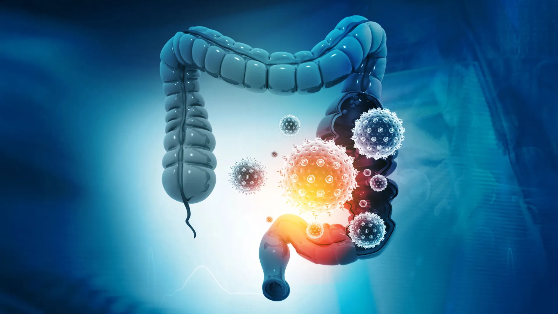
For nearly two decades, scientists have grappled with the insidious mechanism by which a common gut bacterium, Bacteroides fragilis, produces a toxin that inflicts damage upon the colon, thereby contributing to the initiation and progression of tumors. The critical missing piece in this puzzle was the precise manner in which this toxin first latches onto intestinal cells to launch its assault. Now, in a significant scientific breakthrough, researchers have successfully identified this elusive initial step, revealing a surprising vulnerability that could revolutionize approaches to protecting the colon and preventing associated diseases.
The pivotal discovery originates from a collaborative team spearheaded by scientists at the Johns Hopkins Kimmel Cancer Center, its Bloomberg~Kimmel Institute for Cancer Immunotherapy, and the Johns Hopkins University School of Medicine. Their groundbreaking findings, meticulously detailed in the prestigious journal Nature, pinpoint a protein known as claudin-4 as the essential "docking station" or receptor for the bacterial toxin, B. fragilis toxin (BFT). This identification not only unravels a long-standing mystery but also immediately opens pathways for innovative therapeutic interventions. Indeed, the research team has already leveraged this insight to develop a molecular decoy capable of intercepting the toxin before it can inflict damage on delicate colon cells. This novel strategy demonstrated remarkable success in safeguarding mice from the toxin’s deleterious effects, offering a compelling new paradigm for preventing bacterial damage intimately linked with colorectal cancer. The National Institutes of Health provided partial support for this critical investigation.
"We’ve made several attempts over time to identify the receptor, so this is an exciting moment for our team and the broader scientific community," stated Dr. Cynthia Sears, Bloomberg~Kimmel Professor of Cancer Immunotherapy and professor of medicine at Johns Hopkins, and a senior author of the study. Dr. Sears underscored the profound implications of this discovery: "Understanding how bacterial toxins work can open doors to new approaches for detection and therapy for associated diseases, including chronic diarrhea, bloodstream infections, and critically, colorectal cancer."
The Persistent Threat of Bacteroides fragilis and Colorectal Cancer
Colorectal cancer (CRC) remains a significant global health challenge, ranking as the third most common cancer diagnosed in both men and women and the second leading cause of cancer-related deaths worldwide. In the United States alone, the American Cancer Society estimates over 150,000 new cases and more than 50,000 deaths annually. While genetics, diet, and lifestyle are well-established risk factors, mounting evidence increasingly points to the profound influence of the gut microbiome in CRC development.
The bacterium at the heart of this investigation, Bacteroides fragilis, is a common inhabitant of the human gastrointestinal tract, found in the intestines of up to 20% of healthy individuals. While many strains of B. fragilis are benign or even beneficial commensals, a specific subset, known as enterotoxigenic Bacteroides fragilis (ETBF), produces the potent B. fragilis toxin (BFT). It is the presence of these toxin-producing strains, not the bacterium itself, that poses a significant health risk.
The journey to understanding BFT’s role in colon cancer has been a long one, spanning nearly two decades. A landmark 2009 study, also published in Nature Medicine by Dr. Sears and her colleagues, provided compelling evidence that BFT could actively promote the development of colon tumors in animal models. This seminal work was instrumental in establishing a crucial link between specific toxin-producing gut bacteria, chronic inflammation within the colon, and the eventual progression to colorectal cancer. This initial finding ignited intensive research efforts to decipher the toxin’s precise molecular mechanism.
Previous experiments had already elucidated part of BFT’s destructive pathway: the toxin was known to damage a vital protein called E-cadherin. E-cadherin plays a critical role in maintaining the structural integrity of the colon’s protective lining by acting as molecular "glue" that holds neighboring cells together. When BFT cleaves and degrades E-cadherin, this essential barrier becomes compromised, leading to increased permeability of the intestinal wall. This breach can allow harmful substances, pathogens, and inflammatory molecules to penetrate the underlying tissue, fueling chronic inflammation and creating an environment conducive to tumor initiation and growth. However, a critical piece of the puzzle remained stubbornly elusive: despite its ability to dismantle E-cadherin, BFT did not appear to bind directly to it. Researchers hypothesized that an intermediary molecule, an unknown receptor, was facilitating the toxin’s initial attachment to colon cells, guiding it to its ultimate target. Repeated attempts over many years to identify this "missing link" had, until now, met with frustration.
Unmasking the Receptor: A CRISPR-Powered Breakthrough
To finally solve this enduring mystery, the research team turned to CRISPR, a revolutionary gene-editing technology that has transformed biomedical research. CRISPR allows scientists to precisely and systematically disable individual genes within cells, enabling them to observe the downstream effects and infer the function of those genes. This powerful tool offered an unprecedented opportunity to identify genes essential for BFT’s cellular entry and destructive action.
Maxwell White, an M.D./Ph.D. candidate working in Dr. Sears’ laboratory, spearheaded a comprehensive genome-wide CRISPR screening effort. This ambitious project involved a close collaboration with Dr. Matthew Waldor’s laboratory at Harvard Medical School. The methodology involved systematically deactivating genes in colon epithelial cells – the specialized cells that form the inner protective lining of the colon – and then exposing these modified cells to BFT. The goal was to identify which gene disruptions rendered the cells resistant to the toxin’s binding and damage.
Among the thousands of genes screened, one gene stood out with remarkable clarity and consistency: the gene responsible for producing claudin-4. Claudin-4 is a protein integral to the formation of "tight junctions," which are specialized intercellular structures that create a seal between neighboring cells. These tight junctions are crucial for maintaining the intestinal barrier, regulating the selective passage of substances, and preventing harmful elements from leaking into the underlying tissues.
The discovery was striking: when the researchers disabled the gene encoding claudin-4 in colon cells, BFT was no longer able to bind to them. Without this crucial initial attachment point, the toxin was rendered inert, unable to initiate its cascade of damage, specifically its attack on E-cadherin. This definitive finding established claudin-4 as the long-sought receptor for the bacterial toxin, revealing that BFT must first latch onto this protein before it can execute its destructive enzymatic activity. "It took a while to get the assay working and validate the approach, but once we were able to do the screen, claudin-4 was a clear, resounding top hit," Maxwell White recounted, reflecting on the "exciting moment" of discovery.
An Unexpected Target: Redefining Bacterial Toxin Mechanisms
The identity of the BFT receptor came as a significant surprise to the scientific team. Prior hypotheses, based on the known mechanisms of many bacterial toxins, had largely centered on proteins involved in cellular signaling, such as G protein-coupled receptors. These receptors typically act as sophisticated communication switches, enabling cells to perceive and respond to signals from their external environment, triggering complex intracellular pathways.
Claudin-4, however, serves a fundamentally different biological purpose. Its primary role is not in signaling but in the physical architecture and integrity of cell-to-cell connections, specifically within tight junctions. This unexpected discovery revealed a novel and previously uncharacterized strategy for a bacterial toxin to operate. BFT belongs to a class of enzymes known as proteases, which function by cleaving or cutting other proteins. While most proteases directly engage with their targets, BFT appears to necessitate an independent receptor for initial binding before it can access and degrade its ultimate substrate, E-cadherin. A thorough review of existing scientific literature failed to identify another bacterial toxin known to employ this precise two-step mechanism.
To unequivocally confirm the direct physical interaction between BFT and claudin-4, the Johns Hopkins researchers collaborated with structural biologists F. Xavier Gomis-Rüth and Ulrich Eckhard at the Molecular Biology Institute of Barcelona. Utilizing advanced techniques designed to measure molecular interactions, Maxwell White and the Barcelona team provided compelling evidence that BFT and claudin-4 form a stable, tightly bound complex in controlled laboratory settings. These experiments demonstrated a clear one-to-one pairing, with each toxin molecule associating with a single receptor molecule, thereby providing direct physical validation of their interaction.
A Clever Molecular Decoy: Intercepting the Threat
The identification of claudin-4 as the BFT receptor was more than just an academic achievement; it immediately sparked an innovative idea for intervention. If BFT requires claudin-4 to attach to colon cells, then perhaps a cleverly designed substitute could divert the toxin, drawing it away from its intended biological target. This concept formed the basis for developing a novel therapeutic strategy: a molecular decoy.
The team engineered a soluble protein that incorporated specific sequences derived from claudin-4. This "decoy" was meticulously designed to mimic the natural receptor, thereby attracting and binding BFT without allowing the toxin to engage with the actual claudin-4 proteins on the colon’s protective lining. The rationale was simple yet elegant: by saturating the environment with decoy receptors, the toxin would preferentially bind to these harmless stand-ins, effectively neutralizing its ability to initiate cellular damage.
To test the efficacy of this ingenious approach, the researchers collaborated with Dr. Min Dong’s laboratory at Harvard Medical School, specifically working with Kang Wang and other colleagues. These experiments were conducted in living mouse models, providing a more complex and physiologically relevant environment to observe the toxin’s behavior and the decoy’s protective effects within an intact intestinal system.
The results from the mouse studies were highly encouraging and validated the decoy strategy. Instead of binding to the native claudin-4 receptors on the surface of colon cells, BFT preferentially attached to the introduced decoy proteins. By effectively "intercepting" the toxin in this manner, the molecular decoys successfully protected the mice from BFT-induced damage to their colon lining. This finding strongly suggests that it is possible to interfere with the very first step of the toxin’s harmful action, offering a proactive approach to prevention rather than merely attempting to repair the damage after it has occurred.
"This approach could be iterated upon with small molecules or other biologics that have better pharmacological properties," noted Maxwell White, hinting at the future development potential. The research team is now actively exploring various molecular approaches to optimize and enhance the toxin-blocking strategy. This could involve developing small-molecule compounds, which are often amenable to oral administration and exhibit favorable pharmacological profiles, or further refining biologics – treatments derived from biological materials like proteins – to improve their efficacy, stability, and delivery within the human body. Future iterations of the decoy strategy could be engineered for improved performance and ease of administration, moving closer to clinical application.
Implications for Colorectal Cancer Prevention and Beyond
While the discovery of claudin-4 as the BFT receptor and the success of the molecular decoy in mice represent a monumental leap forward, important scientific and clinical questions remain. One key area for future investigation is the precise three-dimensional structure of the BFT-claudin-4 complex. Although advanced artificial intelligence tools, such as AlphaFold, have revolutionized protein structure prediction, they have not yet fully resolved the intricate details of this specific interaction. A definitive understanding of how these two molecules fit together at an atomic level would be invaluable for rationally designing even more effective compounds capable of preventing the toxin from binding. Such structural insights could guide the development of highly specific and potent inhibitors.
Furthermore, extensive additional experiments will be required to determine whether the molecular decoy approach can be safely and effectively translated to humans for the prevention of toxin-related diseases. While the mouse experiments conclusively demonstrated protection against tissue damage, they did not definitively establish that the strategy prevents the long-term development of colorectal cancer. Human clinical trials would be necessary to assess safety, dosage, efficacy, and long-term outcomes.
Nevertheless, this breakthrough definitively resolves a major scientific question that has persisted since the pivotal connection between BFT and colon tumor formation was first established in 2009. By identifying the toxin’s previously unknown point of attachment, researchers have uncovered a powerful new leverage point to interrupt a harmful biological process at its very inception. This fundamental understanding opens the door to a new generation of strategies for detecting, preventing, and treating a range of conditions associated with bacterial toxins. Beyond colorectal cancer, these include chronic diarrheal diseases, where BFT is a known causative agent, and potentially certain bloodstream infections that originate from gut dysbiosis and barrier compromise. The ability to neutralize this specific bacterial toxin holds immense promise for improving human health.
Study Authors, Funding, and Disclosures
The comprehensive study benefited from the dedicated contributions of numerous researchers. In addition to Dr. Cynthia Sears and Maxwell White, key contributors from Johns Hopkins included Jason Chen, Shaoguang Wu, Abby L. Geis, and Jessica Queen. Collaborators from Harvard Medical School included Hailong Zhang, Karthik Hullahalli, and Jie Zhang, along with Kang Wang and colleagues from Dr. Min Dong’s laboratory. The structural biology aspects were critically supported by F. Xavier Gomis-Rüth and Ulrich Eckhard at the Molecular Biology Institute of Barcelona.
The research received generous financial support from several prestigious organizations, including the Bloomberg~Kimmel Institute for Cancer Immunotherapy, Janssen Research and Development, Cancer Research UK, and the National Institutes of Health (under grant numbers R01 AI042347, R01 NS080833, R01 NS117626, R01 AI170835, and R01 AI189789). Additional funding was provided by the Howard Hughes Medical Institute. These collaborations and funding mechanisms underscore the interdisciplinary and international nature of modern scientific discovery, pooling expertise and resources to tackle complex biomedical challenges.
