
 By Nana O
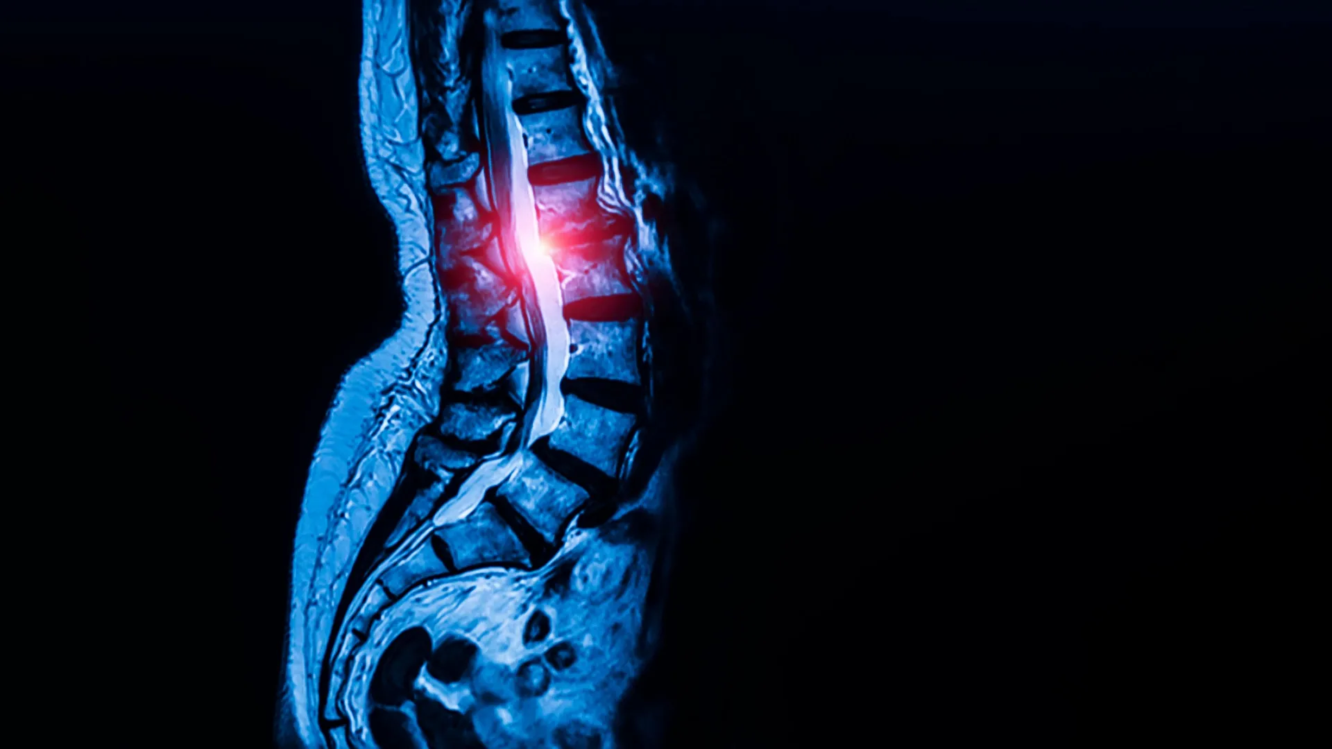
New York, NY – In a groundbreaking discovery poised to reshape our understanding of connective tissue health and disease, researchers at Weill Cornell Medicine and the Hospital for Special Surgery (HSS) have pinpointed a previously elusive population of stem cells. These specialized cells, identified through meticulous research, appear to be the sole architects responsible for generating the body’s tendons and ligaments—the vital connective tissues that anchor muscles to bones and stabilize joints. This landmark finding not only illuminates the fundamental mechanisms of musculoskeletal development but also presents a promising new avenue for treating debilitating conditions like lumbar spinal stenosis, a widespread ailment affecting millions globally.
The study, published on September 7th in the prestigious journal Cell, details how scientists successfully isolated and characterized these unique stem cells. Their investigation went further, uncovering compelling evidence that an overactivity of these very cells in the lower spine may be a significant contributor to the development of lumbar spinal stenosis. This condition, which impacts an estimated 103 million people worldwide, occurs when the ligaments within the spinal canal thicken, progressively narrowing the space available for the spinal cord and nerves. This compression can lead to a cascade of debilitating symptoms, including chronic pain, persistent numbness, and significant difficulties with mobility, profoundly impacting the quality of life for affected individuals.
The implications of this discovery are profound, offering the tantalizing possibility of developing novel therapeutic strategies that target these newly identified stem cells. One particularly exciting prospect involves exploring the potential of existing medications, such as those currently prescribed to manage high blood pressure, as a treatment modality for spinal stenosis.
"For years, the scientific community has debated the precise cellular origins of tendons and ligaments," stated Dr. Matthew Greenblatt, co-corresponding author of the study, the Rohr Family Research Scholar, and an associate professor of pathology and laboratory medicine at Weill Cornell. "While various candidate stem cell populations had been proposed, none had definitively demonstrated the capacity to both self-renew and generate the full diversity of mature cell types found in tendons and ligaments. Our work finally provides that definitive answer."
Following their successful identification of these specialized stem cells, the research team pivoted to investigate their potential role in the pathogenesis of lumbar spinal stenosis. This focus was driven by the urgent need for improved treatment options for patients suffering from this condition, particularly those whose symptoms have progressed to a severity where surgery is often the only recourse.
Dr. Sravisht Iyer, co-corresponding author and an associate professor of orthopedics at Weill Cornell and a spine surgeon at HSS, emphasized the transformative potential of this research. "Identifying these specialized stem cells opens up an entirely new frontier in research, allowing us to approach this disease with a mechanistic understanding rather than simply managing symptoms as they worsen and necessitate surgical intervention to alleviate nerve compression," he explained. "These findings are incredibly exciting because they hold the promise of fundamentally changing how we deliver spinal care."
Unraveling the Mystery: The Quest for Tendon and Ligament Stem Cells
The journey to identify the stem cells responsible for tendon and ligament formation was a complex and protracted one, building upon years of prior research by Dr. Greenblatt and his colleagues. The team had previously made significant strides in mapping the landscape of skeletal stem cells, including the identification of the crucial stem cell population that initiates fracture repair in the outer layer of bone in 2018. Subsequent research by the group also shed light on the stem cells involved in the formation of the skull and spine.
However, isolating a comparable stem cell for tendons and ligaments proved to be a far more formidable challenge. Unlike bone, which has a more defined cellular structure, tendons and ligaments are composed of a dense network of fibroblast-like cells that exhibit a high degree of similarity in appearance. This morphological resemblance made it exceedingly difficult to distinguish one specific stem cell population from the multitude of other cells present in these tissues.
"We embarked on an exhaustive process, analyzing thousands upon thousands of individual cells," Dr. Greenblatt recounted. "Our approach involved meticulously sorting these cells into distinct types and then rigorously identifying which population possessed the defining characteristics of ‘stemness’."
The concept of "stemness" in this context refers to the inherent ability of a rare cell population to not only continually replicate itself, thereby maintaining a reservoir of progenitor cells, but also to differentiate into the mature, specialized cell types required for the formation and ongoing maintenance of tendon and ligament tissue. Initial investigations in mouse models provided crucial insights. The researchers discovered these stem cells residing within a specialized niche located within the tendons and ligaments themselves. This anatomical localization suggests that these niches act as critical reservoirs, essential for tissue growth, repair, and regeneration throughout the lifespan of an organism.
Leveraging the knowledge gained from their mouse studies, the team then applied their findings to search for analogous cell populations in human tissues. Human ligament samples were ethically obtained from tissue that had been surgically removed by Dr. Iyer from consenting patients. This critical step allowed for direct comparison and validation of the cellular mechanisms observed in animal models within the human biological context.
The research, spearheaded by first author Dr. Lingling Hu, a postdoctoral fellow in both Dr. Greenblatt’s and Dr. Iyer’s laboratories, ultimately confirmed the presence of these specialized stem cells in humans. The human cells demonstrated the same dual capacity observed in mice: the ability to self-renew and the potential to generate mature ligament cells.
Furthermore, the study revealed that these stem cells were not confined to a single anatomical location. Their presence extended far beyond the spine, indicating a systemic role in connective tissue development.
"Our investigations spanned across various anatomical sites," Dr. Greenblatt confirmed. "We examined the patellar ligament in the kneecap, and we analyzed the Achilles tendon. In every location we investigated, we found this remarkable cell. This leads us to believe that we have identified the universal stem cell responsible for the formation and maintenance of tendons and ligaments throughout the entire body."
The Link to Spinal Stenosis: Uncontrolled Growth in the Spine
The research team then turned their attention to exploring the potential involvement of these newly identified stem cells in the development of lumbar spinal stenosis. To this end, they compared stem cells harvested from patients diagnosed with spinal stenosis against stem cells collected from the spinal ligaments of individuals who had undergone surgery for herniated discs but exhibited no signs of stenosis. These human samples were also obtained with informed consent.
The findings were striking: ligaments from patients with spinal stenosis contained a significantly higher abundance of the newly identified stem cells compared to those from healthy individuals. To further elucidate the functional implications of this increased cell population, the researchers transplanted these stem cells into a mouse model. The results showed that stem cells derived from stenotic ligaments produced a greater quantity of tendon cells than stem cells obtained from individuals without stenosis.
"While lumbar spinal stenosis is recognized as a complex multifactorial condition, our findings provide compelling evidence that these specific stem cells play a direct role in the pathology," Dr. Greenblatt asserted. Dr. Greenblatt is also an integral member of the Sandra and Edward Meyer Cancer Center at Weill Cornell, a testament to his broad research interests in cellular mechanisms.
Delving deeper into the cellular mechanisms at play, the researchers observed a distinct difference in the signaling pathways within the stem cells associated with spinal stenosis. Specifically, these cells exhibited heightened calcium signaling activity when compared to their healthy counterparts. Calcium signaling serves as a critical intracellular communication system, regulating a wide array of cellular processes, including growth, proliferation, and metabolic activity.
Intriguingly, when the scientists experimentally increased calcium signaling in stem cells derived from healthy ligaments, they were able to induce an uncontrolled and excessive growth of tissue, mimicking the pathological changes seen in spinal stenosis. This experiment provided a direct causal link between elevated calcium signaling and abnormal connective tissue proliferation.
A Potential Therapeutic Target: Leveraging Existing Medications
The converse observation proved equally significant. When the researchers deliberately reduced calcium signaling in a mouse model engineered to exhibit lumbar spinal stenosis, the abnormal cell growth was effectively halted. This crucial finding strongly suggests that calcium signaling represents a viable therapeutic target for interventions aimed at treating spinal stenosis.
Furthermore, this discovery opens up the exciting possibility of repurposing existing pharmacological agents. Calcium channel blockers, a well-established class of drugs widely employed in the management of hypertension (high blood pressure), function by modulating calcium signaling. The research team hypothesizes that these readily available medications could potentially be investigated as a novel treatment strategy for spinal stenosis. However, extensive clinical trials will be imperative to rigorously assess the safety and efficacy of such an approach in human patients before any widespread clinical application can be considered.
"This research likely represents the very first work to identify a potential therapeutic target for one of the most prevalent spine conditions globally," Dr. Iyer remarked. "The potential to move beyond palliative care and address the underlying cellular mechanisms of spinal stenosis is truly revolutionary."
The ramifications of this discovery are likely to extend beyond the immediate scope of spinal stenosis. Dr. Greenblatt has articulated plans to investigate whether these same tendon and ligament stem cells are implicated in other connective tissue disorders. Conditions such as Marfan syndrome, a genetic disorder that profoundly affects connective tissues throughout the body, are prime candidates for future exploration.
This breakthrough could also provide invaluable insights into why certain tendon and ligament injuries prove exceptionally difficult to heal. Areas of future research may include understanding the cellular basis of chronic conditions like rotator cuff tears, Achilles tendon injuries, the challenges associated with ligament reconstruction, and the mechanisms underlying chronic tendon degeneration.
"Given that this cell appears to be the ultimate progenitor of all tendon and ligament cells, it is highly probable that defects or dysregulation within this cell population are at the root of a wide spectrum of tendon and ligament disorders," Dr. Greenblatt concluded.
This extensive research was made possible through significant funding from a variety of sources, including the Marfan Foundation’s Victor A. McKusick Fellowship, a Kellen Scholars Award, the Children’s Tumor Foundation (grant CTF-2023-01-005), the Arthritis National Research Foundation (grant 1065843), a Jumpstart award from Weill Cornell, a National Research Foundation of Korea (NRF) award funded by the Ministry of Education (grant NRF-2021R1A6A3A14038667), the National Institutes of Health (grants T32-AR078751 and T32-AR071302-07), a MIND Prize from the Pershing Square Foundation, a Mary Kay Ash Foundation Award, an Innovator Award from the Marfan Foundation, and a Burroughs Wellcome Career Award for Medical Scientists. The collaborative effort underscores the critical role of sustained investment in fundamental scientific inquiry to drive medical innovation.
