
 By George Ongkojoyo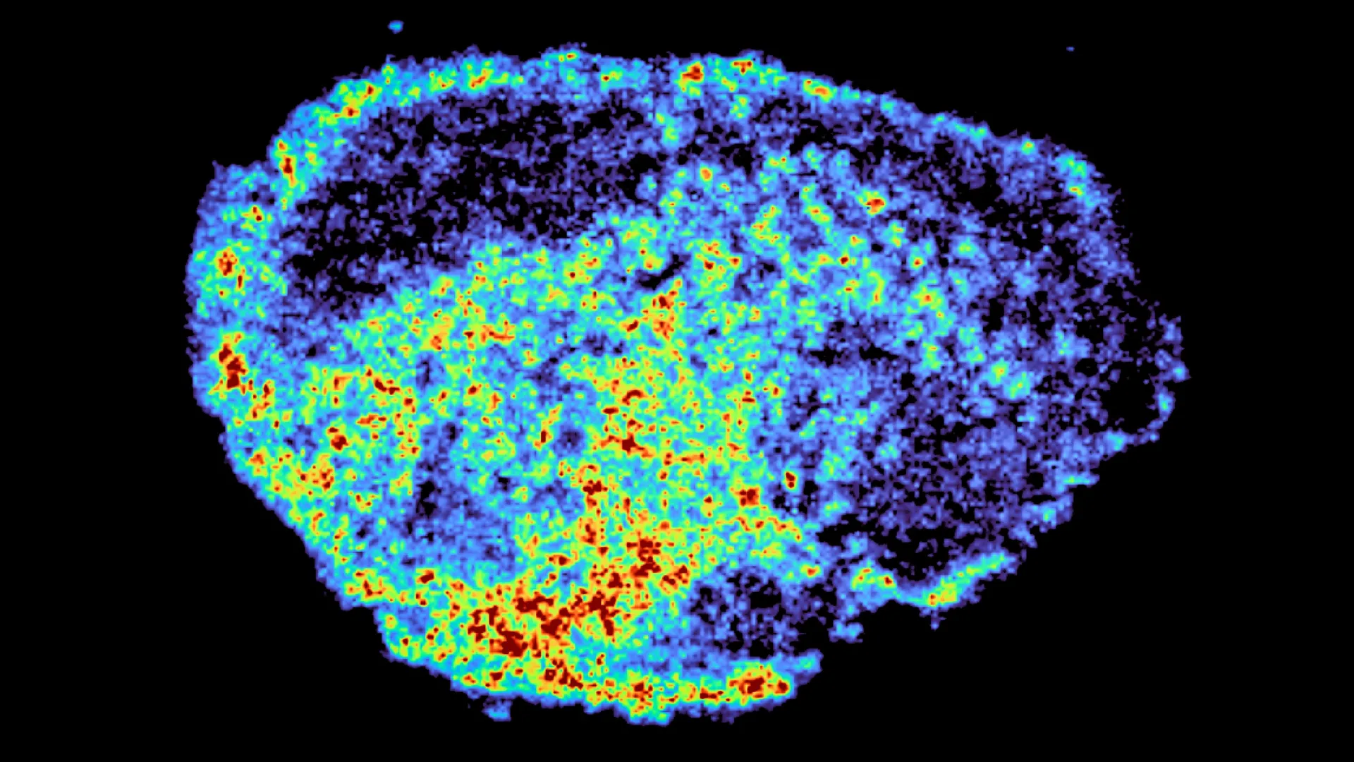
The landscape of modern oncology is defined by a central paradox: while targeted therapies have revolutionized survival rates for millions, the efficacy of these treatments remains frustratingly inconsistent across patient populations. A landmark study recently published in the journal Nature Communications has provided a critical breakthrough in understanding this phenomenon. Led by Dr. Louise Fets at the MRC Laboratory of Medical Sciences (LMS), a multidisciplinary team of researchers has identified a previously overlooked mechanism within cancer cells that dictates how drugs are distributed, stored, and ultimately utilized. By focusing on PARP inhibitors—a cornerstone of ovarian cancer treatment—the study reveals that the internal architecture of a cell, specifically the role of lysosomes, acts as a decisive factor in whether a therapy succeeds or fails.
The Evolution and Challenge of PARP Inhibitors in Clinical Oncology
Poly (ADP-ribose) polymerase (PARP) inhibitors represent one of the most significant advancements in precision medicine over the last two decades. Designed to exploit the vulnerabilities of cancer cells with defective DNA repair mechanisms—most notably those with BRCA1 or BRCA2 mutations—these drugs function by preventing cancer cells from repairing single-strand DNA breaks. When these breaks persist, they evolve into double-strand breaks during replication, which a BRCA-deficient cell cannot fix, leading to programmed cell death.
Since the first PARP inhibitor received regulatory approval, the class has expanded to include several key agents: olaparib, rucaparib, niraparib, and talazoparib. While these drugs have transformed the prognosis for high-grade serous ovarian cancer, clinical data indicates a significant spectrum of response. Some patients achieve long-term remission, while others exhibit primary resistance or develop secondary resistance after an initial period of efficacy. Traditionally, researchers attributed this resistance to genetic mutations or the upregulation of "efflux pumps" that eject drugs from the cell. However, the new findings from the MRC Laboratory of Medical Sciences suggest that the physical distribution of the drug within the tumor microenvironment is equally, if not more, influential.
Methodological Innovation: Explants and Spatial Mapping
To investigate the internal dynamics of drug distribution, Dr. Fets and her colleagues moved away from traditional two-dimensional cell cultures, which often fail to replicate the complex 3D architecture of human tumors. Instead, the team utilized "explants"—thin, live slices of ovarian tumors donated by patients. These samples were maintained in a laboratory environment that preserved their structural integrity and cellular diversity, providing a high-fidelity model of how drugs interact with real human tissue.
The researchers employed a sophisticated dual-imaging approach to track the movement of PARP inhibitors. The first component involved mass spectrometry imaging (MSI), a powerful tool that allows scientists to visualize the exact location of drug molecules within a tissue sample by measuring their molecular weight. By overlaying these drug maps with spatial transcriptomics—a method that measures gene activity across different regions of the same tissue—the team could correlate high and low drug concentrations with specific cellular behaviors and genetic expressions.
This methodology revealed a startling level of heterogeneity. Even within a single tumor sample treated with a uniform dose of a PARP inhibitor, some regions were saturated with the drug while neighboring areas remained virtually untouched. This uneven spread suggests that the "average" concentration of a drug in a tumor, a metric often used in clinical trials, may be a misleading indicator of actual therapeutic coverage.
The Lysosome: From Waste Center to Drug Reservoir
The most significant discovery of the study involves the role of lysosomes. Historically viewed as the "recycling centers" or "garbage disposals" of the cell, lysosomes are acidic organelles responsible for breaking down cellular waste. The research team found that certain PARP inhibitors are chemically drawn into these acidic compartments.
Once inside the lysosome, these drugs become "trapped." However, this sequestration is not necessarily a negative outcome. The study found that lysosomes act as slow-release reservoirs. They accumulate high concentrations of the drug and then gradually release it back into the rest of the cell over time. This process effectively extends the duration of the cell’s exposure to the medication, potentially enhancing its lethal effect on the cancer.
"We were surprised to see large variability in drug accumulation at the single-cell level," noted Dr. Carmen Ramirez Moncayo, the study’s first author and a Postdoctoral Researcher at the LMS. "This variability was driven by the build-up of a drug in lysosomes, which are acting as reservoirs, increasing the exposure of cancer cells to drugs by storing and releasing the drug when needed."
Crucially, the study highlighted that not all drugs in this class interact with lysosomes in the same way. The researchers observed that rucaparib and niraparib were significantly affected by lysosomal trapping, whereas olaparib was not. This distinction is likely due to the specific chemical properties of the molecules, such as their lipophilicity and basicity, which influence how they cross organelle membranes.
Comparative Data and Spatial Transcriptomics Findings
The integration of spatial transcriptomics allowed the team to go a step further than mere visualization. By examining the RNA profiles of cells in drug-rich versus drug-poor areas, the researchers could see how the cells were reacting to the treatment in real-time.
The data indicated that cells with high lysosomal drug accumulation showed stronger signatures of DNA damage and stress, confirming that the "reservoir effect" was functionally active. Conversely, areas with low drug penetration showed active survival signaling, providing a potential roadmap for how tumors "nest" resistant cells in pockets where the drug cannot reach effective concentrations.
Dr. Zoe Hall, senior author and Associate Professor at Imperial College London’s Department of Metabolism, Digestion and Reproduction, emphasized the importance of this spatial context. "Through the spatial mapping of drug molecules, we could pinpoint regions of high and low drug and compare gene expression from the same tissue slice. This allowed us to see exactly what the cells were doing in response to the specific amount of drug they were receiving," she explained.
Chronology of Research and Institutional Support
The study represents several years of collaborative effort between the MRC Laboratory of Medical Sciences, Imperial College London, and various clinical partners. The project was initiated to address the "black box" of drug pharmacokinetics—the study of how a body affects a drug—at a microscopic level.
The research was supported by a robust network of funding bodies, reflecting its importance in the field of oncology. Contributors included the Medical Research Council (MRC), Cancer Research UK (CRUK), and the Integrative Toxicology Training Partnership. Additionally, the study received a Victoria’s Secret Global Fund for Women’s Cancers Career Development Award, in partnership with Pelotonia and the American Association for Cancer Research (AACR). This diverse funding highlights the global interest in refining ovarian cancer treatments, a disease that remains the leading cause of death from gynecologic cancers in many developed nations.
Implications for Personalized Medicine and Future Drug Design
The implications of these findings for the future of cancer care are profound. If the efficacy of a drug is dependent on how it is stored within lysosomes, then a patient’s "lysosomal profile" could become a vital biomarker. In the future, clinicians might analyze a patient’s tumor not just for genetic mutations like BRCA, but also for the density and activity of lysosomes.
Furthermore, this research provides a new lens through which to view drug resistance. If a patient relapses, it may not be because the cancer has "learned" to ignore the drug, but because the tumor’s physical structure or cellular composition has changed in a way that prevents the drug from accumulating in these essential lysosomal reservoirs.
Dr. Louise Fets, the study’s lead author and Head of the LMS’ Drug Transport and Tumour Metabolism Group, views this as a stepping stone toward a more tailored therapeutic approach. "By understanding how drugs are taken up into cells, we can understand whether this influences why cancer drugs work for some people and not for others. Eventually, we hope to be able to study the molecular signature of a patient’s tumor to help to tailor therapeutic approaches in a more personalized way."
Addressing the Limitations: From Lab to Clinic
While the results from tumor explants are compelling, the researchers acknowledge the challenges of translating these findings into the human body. In a clinical setting, a drug must navigate the patient’s circulatory system before it even reaches the tumor. Cancerous tumors are notorious for having "leaky" and disorganized blood vessels, which creates high interstitial pressure that can block drug delivery.
The next phase of research will involve animal models to study how the systemic delivery of these drugs interacts with the lysosomal trapping observed in this study. Researchers also plan to expand their patient cohorts to include those with relapsed cancers to see if lysosomal behavior changes after repeated exposure to chemotherapy.
Conclusion: A New Frontier in Pharmacological Research
The study published in Nature Communications marks a shift in how scientists think about "targeted" therapy. It suggests that "targeting" a tumor is only half the battle; the other half is ensuring the drug is distributed effectively once it arrives. By identifying lysosomes as hidden drug reservoirs, the MRC Laboratory of Medical Sciences has opened a new door for optimizing existing treatments and designing the next generation of anti-cancer agents. As the medical community moves closer to the ideal of truly personalized medicine, understanding the microscopic geography of a tumor will be essential in ensuring that no patient is left behind by therapies that have the potential to save their lives.
