
 By George Ongkojoyo
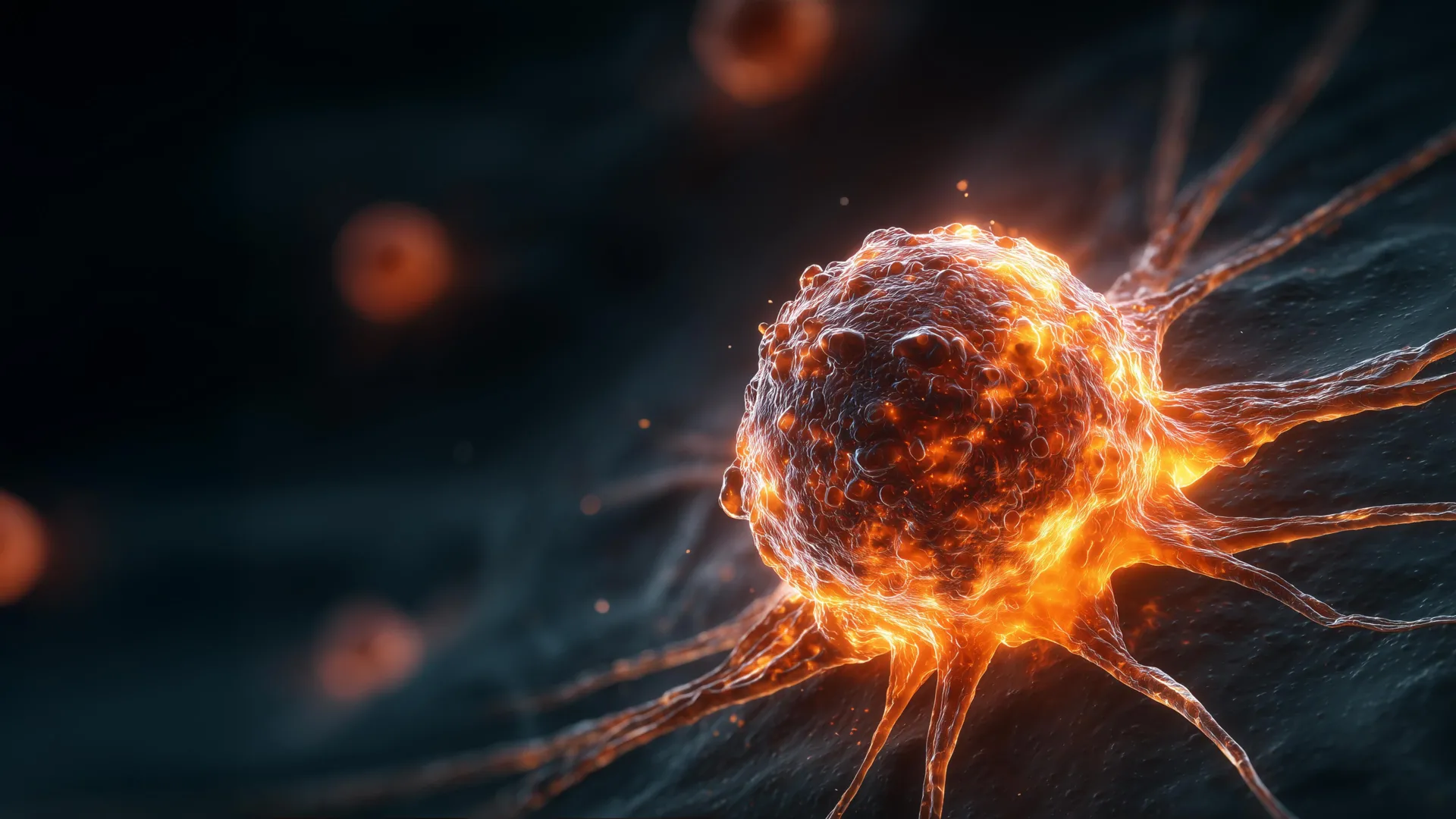
In a series of groundbreaking studies that challenge long-held assumptions in oncology, researchers at Virginia Tech have identified new mechanisms that explain why some cancers are more aggressive and resistant to treatment than others. The findings, led by a team of cell biologists and graduate researchers, suggest that the danger of a tumor is dictated not just by the presence of abnormal cells, but by how those cells manipulate their environment and how their physical size correlates with their survival potential. By examining the behavior of "tetraploid" cells—cells containing double the normal amount of genetic material—the team has uncovered a paradoxical relationship between cell volume and malignancy, while also demonstrating how these abnormal cells recruit healthy tissue to facilitate rapid tumor expansion.
The research, which spanned five years of intensive laboratory investigation, was recently detailed in the Proceedings of the National Academy of Sciences (PNAS), with supplementary findings appearing in the journal Cancer Research. The work provides a significant leap forward in understanding chromosomal instability, a hallmark of many of the most difficult-to-treat human cancers, including breast and colorectal malignancies.
The Microscopic Foundation of Oncology Research
The path to these discoveries began in the highly controlled environment of the Virginia Tech laboratories, where graduate researcher Megan Sweet performs the delicate task of preparing tumor samples for analysis. The process is a blend of high-tech precision and manual craftsmanship. Working within a refrigerated metal chamber known as a cryostat, Sweet prepares sections of tumor tissue grown in mouse models. These tumors, often no larger than a pinkie fingernail, must be sliced into sections so thin that they are translucent, allowing light from a microscope to pass through the cellular structures.
This meticulous preparation is essential for the subsequent staining process, where specific proteins and genetic markers are highlighted to reveal the inner workings of the tumor. "It’s all about fine-tuning and making sure it’s going to be an even slice," Sweet noted during a demonstration of the technique. The rhythmic sound of the microtome blade slicing through the frozen tissue marks the beginning of a process that allows scientists to compare the architectural differences between slow-growing and aggressive tumors. This painstaking work has allowed the team to observe how cells with abnormal chromosome counts interact with their surrounding environment at a granular level.
Understanding Chromosomal Abnormalities: The Tetraploid Factor
To understand the team’s findings, one must first look at the fundamental biology of human cells. Most healthy cells are diploid, containing two sets of chromosomes—one from each parent. During normal cell division, these chromosomes are replicated and then evenly distributed into two new daughter cells. However, this process can fail. When a cell replicates its DNA but fails to divide, it becomes tetraploid, possessing four sets of chromosomes.
While the human body has mechanisms to identify and eliminate such errors, some tetraploid cells survive and continue to divide. This state of "whole genome doubling" is increasingly recognized as a precursor to cancer. It provides a reservoir of genetic material that allows cancer cells to evolve rapidly, developing resistance to chemotherapy and the ability to metastasize. The Virginia Tech team, led by cell biologist Daniela Cimini, sought to determine exactly how these tetraploid cells influence the overall trajectory of a tumor.
The Recruitment Paradox: How Cancer Hijacks Healthy Tissue
One of the most significant findings of the study involves the interaction between tetraploid cancer cells and the host’s healthy tissue. In experiments comparing tumors derived from ordinary diploid cells with those derived from tetraploid cells, researchers Sweet and Mat Bloomfield observed a puzzling phenomenon.
As the tetraploid-derived tumors grew in mouse models, the actual percentage of tetraploid cells within the mass began to decrease. Under traditional logic, a decrease in the most "abnormal" cells should result in a slowing of tumor growth. Instead, the researchers found the opposite: the tumors continued to expand at an accelerated rate.
The investigation revealed that the tetraploid cells were functioning as "recruiters." Even when present in small numbers, these cells triggered the recruitment of stromal cells—non-cancerous connective tissue cells that normally provide structural support to organs. Once recruited into the tumor microenvironment, these healthy cells were effectively "reprogrammed" to support the cancer, providing the structural scaffolding and chemical signals necessary for the tumor to flourish.
"The presence of even a small fraction of these tetraploid cells can promote the recruitment of extra non-cancerous cells that support further tumor progression," Sweet explained. This discovery shifts the focus of cancer research from looking solely at the cancer cells themselves to examining the "neighborhood" the cells create. It suggests that a tumor’s danger is not just a result of its own growth, but its ability to corrupt the healthy biological processes of the host.
Challenging Biological Norms: The Role of Cell Size
Parallel to the study of tissue recruitment, graduate student Mat Bloomfield uncovered a second, equally surprising trend regarding the physical dimensions of cancer cells. In the world of biology, there is a generally accepted "scaling" rule: the more DNA a cell has, the larger it should be. Because tetraploid cells have twice the DNA of diploid cells, they are expected to be roughly twice the size.
However, when Bloomfield established colonies from individual tetraploid cells, he found significant variation. While many followed the expected size rules, a subset of tetraploid cells was 25 to 30 percent smaller than anticipated. When these "small" tetraploid cells were tested, they exhibited far more aggressive traits than their larger counterparts.
The data showed that these smaller cells:
- Divided and grew at a significantly faster rate.
- Demonstrated a higher capacity for invasion into surrounding tissues.
- Showed increased tolerance to common anti-cancer drugs and environmental stressors.
This finding was consistent across different types of cancer, including breast and colorectal models, suggesting a universal biological principle at play. The smaller size may provide a metabolic advantage, allowing the cells to move more easily or maintain a more efficient ratio of surface area to volume, though the exact biological mechanism remains a subject for future study.
Analyzing Human Data: From the Lab to the Clinic
To ensure their laboratory findings were relevant to human health, the Virginia Tech team turned to the Cancer Genome Atlas (TCGA), a landmark program that has cataloged the genetic and physical characteristics of thousands of patient samples across dozens of cancer types.
By analyzing this vast data set, the researchers confirmed that the relationship between cell size and patient outcomes was not limited to mouse models. In human patients, tumors characterized by smaller tetraploid cells were statistically associated with worse prognoses and lower overall survival rates. This correlation held true across multiple cancer types, suggesting that cell size could serve as a powerful new biomarker for oncologists.
"We already knew that tetraploidy can make cells more tumorigenic, but now we know that if you incorporate the size of the cells, it can be more predictive of tumorigenic potential," said Daniela Cimini. This adds a new layer to diagnostic pathology. Currently, pathologists look at cell shape and chromosome counts; the Virginia Tech research suggests that measuring the precise volume of these cells could provide a more accurate forecast of how a patient’s disease will progress.
Implications for Future Oncology and Treatment Strategies
The implications of this research are manifold. First, it highlights the importance of the tumor microenvironment. If the recruitment of healthy stromal cells is a primary driver of aggression in tetraploid-heavy tumors, then therapies designed to block that recruitment—rather than just killing the cancer cells—could be a viable path forward.
Second, the discovery regarding cell size offers a potential new metric for "risk-stratifying" patients. Patients whose biopsies show smaller, high-ploidy cells might require more aggressive initial treatment or more frequent monitoring, as their tumors are more likely to resist standard chemotherapy.
The scientific community has reacted with interest to the Virginia Tech findings. While the link between aneuploidy (abnormal chromosome numbers) and cancer has been studied since the early 20th century, the specific role of cell size and the "recruitment" mechanism provides a modern update to classical theories. By combining high-resolution laboratory work with "big data" analysis of human samples, the team has bridged the gap between basic cell biology and clinical application.
A Continuing Search for Answers
For the researchers at Virginia Tech, the publication of these papers marks the beginning of a new phase of inquiry. The team plans to delve deeper into the molecular signals that tetraploid cells use to communicate with stromal tissue. Identifying the specific proteins or chemical "messages" involved in this recruitment could lead to the development of new drugs designed to "deafen" the healthy cells to the cancer’s commands.
Additionally, the team is working to understand why some tetraploid cells become smaller in the first place. Whether this is a result of rapid, "shrunken" divisions or a specific metabolic adaptation remains to be seen.
As Megan Sweet continues her work at the microtome, the rhythmic sound of the blade serves as a reminder of the steady, incremental progress of science. Each thin slice of tissue brings the team closer to understanding the complex, often deceptive strategies that cancer uses to survive. In the fight against a disease that evolves as quickly as cancer, the discovery that size and recruitment matter as much as genetics provides a vital new map for researchers and clinicians alike.
