
 By Nana O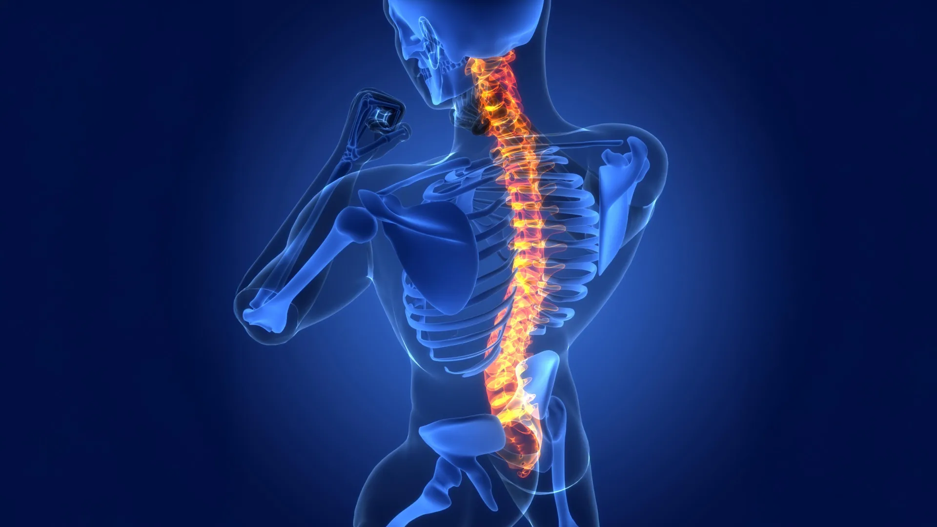
Researchers at Cedars-Sinai have made a groundbreaking discovery, identifying a previously unknown biological repair process within the central nervous system that holds significant promise for future treatments of spinal cord injuries, stroke, and neurodegenerative diseases such as multiple sclerosis. The findings, meticulously detailed in the prestigious scientific journal Nature, reveal an unexpected and crucial role for astrocytes, a fundamental type of glial cell long understood as support staff for neurons, in orchestrating tissue repair from a distance. This revelation challenges previous assumptions about glial cell function and opens new avenues for therapeutic intervention.
Astrocytes: Beyond Support, Towards Active Repair
"Astrocytes are critical responders to disease and disorders of the central nervous system — the brain and spinal cord," stated neuroscientist Joshua Burda, PhD, assistant professor of Biomedical Sciences and Neurology at Cedars-Sinai and senior author of the study. "We discovered that astrocytes far from the site of an injury actually help drive spinal cord repair. Our research also uncovered a mechanism used by these unique astrocytes to signal the immune system to clean up debris resulting from the injury, which is a critical step in the tissue-healing process."
The research team has aptly named these crucial distant-acting cells "lesion-remote astrocytes," or LRAs. Further investigation revealed that these LRAs are not a monolithic group but comprise several distinct subtypes, each with specialized functions. For the first time, this study elucidates how one specific subtype of LRA can detect damage occurring elsewhere in the central nervous system and initiate a cascade of responses essential for recovery.
Understanding Spinal Cord Injury: A Complex Landscape
To fully appreciate the significance of this discovery, it is important to understand the anatomy and vulnerability of the spinal cord. The spinal cord is a vital, long bundle of nerve tissue that acts as the primary communication highway between the brain and the rest of the body, extending from the base of the skull down the vertebral column. Its internal structure is organized into two main regions: the inner gray matter, characterized by its butterfly or H-shape, which contains nerve cell bodies (neurons), glial cells including astrocytes, and synapses; and the surrounding white matter, composed primarily of myelinated nerve fibers (axons) bundled together, along with astrocytes. These myelinated axons are responsible for transmitting electrical signals rapidly over long distances, enabling complex motor commands, sensory perception, and autonomic regulation.
Astrocytes, historically viewed as passive structural and metabolic support for neurons, play an indispensable role in maintaining the delicate electrochemical balance and structural integrity of the central nervous system (CNS). They provide crucial nutrients, regulate the extracellular environment by controlling ion concentrations and neurotransmitter levels, contribute to the blood-brain barrier, and are involved in synaptic function and plasticity.
When the spinal cord sustains an injury, whether through trauma, stroke, or the progression of a disease, the consequences can be devastating. Nerve fibers are often severed or severely damaged, leading to a loss of communication between the brain and the body. This disruption can manifest as paralysis, loss of sensation, and autonomic dysfunction. A significant challenge in spinal cord injury is the subsequent breakdown of damaged nerve fibers into cellular debris. In most peripheral tissues, inflammation is a localized response to injury, helping to clear pathogens and initiate repair. However, within the CNS, the long-ranging nature of nerve fibers means that damage and the subsequent inflammatory response can spread far beyond the initial injury site, potentially causing secondary damage and hindering recovery. The accumulation of fatty debris from degenerating myelin sheaths, a protective covering on axons, can further exacerbate inflammation and impede the natural healing processes.
The Unveiling of Lesion-Remote Astrocytes (LRAs)
The Cedars-Sinai research team conducted a series of rigorous experiments utilizing mouse models with induced spinal cord injuries. Their observations provided compelling evidence that LRAs are not merely bystanders but active participants in promoting repair. Crucially, the study also identified strong indicators that this same CCN1-mediated repair process is operative in human spinal cord tissue samples from patients, suggesting a conserved and evolutionarily significant mechanism.
The CCN1 Signaling Pathway: A Key to Immune System Coordination
A pivotal finding of the study is the identification of a specific subtype of LRA that produces a protein called CCN1. This molecule, a member of the cysteine-rich protein family, acts as a potent signaling factor, directing the activity of resident immune cells in the CNS, known as microglia.
"One function of microglia is to serve as chief garbage collectors in the central nervous system," explained Dr. Burda. "After tissue damage, they eat up pieces of nerve fiber debris — which are very fatty and can cause them to get a kind of indigestion. Our experiments showed that astrocyte CCN1 signals the microglia to change their metabolism so they can better digest all that fat."
This enhancement of microglial metabolic efficiency, driven by astrocyte-derived CCN1, is a critical step in clearing the inflammatory milieu and creating a more permissive environment for tissue regeneration. According to Dr. Burda, this improved debris removal may offer a biological explanation for why some patients experience partial, spontaneous recovery after spinal cord injury. Conversely, when the researchers experimentally eliminated or blocked the production of astrocyte-derived CCN1, the healing process was significantly impaired.
"If we remove astrocyte CCN1, the microglia eat, but they don’t digest," Dr. Burda elaborated. "They call in more microglia, which also eat but don’t digest. Big clusters of debris-filled microglia form, heightening inflammation up and down the spinal cord. And when that happens, the tissue doesn’t repair as well." This highlights the delicate balance required for effective CNS repair and underscores the detrimental effects of unchecked inflammation and inefficient debris clearance.
Broader Implications: Multiple Sclerosis, Stroke, and Beyond
The implications of this discovery extend far beyond spinal cord injuries. The research team further investigated spinal cord tissue samples from individuals diagnosed with multiple sclerosis (MS), a chronic autoimmune disease characterized by inflammation and demyelination in the CNS. Their analysis revealed the presence of the same CCN1-related repair process, suggesting that LRAs and their signaling mechanisms may play a role in mitigating the chronic inflammation and tissue damage characteristic of MS.
"The role of astrocytes in central nervous system healing is remarkably understudied," commented David Underhill, PhD, chair of the Department of Biomedical Sciences at Cedars-Sinai. "This work strongly suggests that lesion-remote astrocytes offer a viable path for limiting chronic inflammation, enhancing functionally meaningful regeneration, and promoting neurological recovery after brain and spinal cord injury and in disease."
The findings also hold significant relevance for stroke recovery. Strokes, caused by interruption of blood supply to the brain, result in neuronal cell death and subsequent tissue damage, often accompanied by inflammatory responses. Understanding how astrocytes at a distance can orchestrate immune cell activity to clear debris and promote a healing environment could be pivotal in developing new neuroprotective and restorative strategies for stroke patients.
A Timeline of Discovery and Future Directions
The path to this discovery involved years of dedicated research, building upon foundational knowledge of glial cell biology and neuroinflammation. While the precise timeline of the research project leading to the Nature publication is not detailed in the provided text, typical scientific breakthroughs of this magnitude often involve iterative phases of hypothesis generation, experimental design, data acquisition, rigorous analysis, and peer review.
- Early Hypotheses and Observations: Researchers likely began with observations of astrocytic involvement in CNS injury and disease, perhaps noticing their presence in areas surrounding lesions or in response to inflammatory signals. Initial investigations might have focused on astrocytes immediately adjacent to damaged areas.
- Identifying the "Remote" Component: A critical turning point would have been the realization that astrocytes located significantly away from the injury site were also actively involved, leading to the conceptualization of "lesion-remote astrocytes."
- Mechanism Elucidation: The focus would then shift to understanding how these remote astrocytes exert their influence. This phase would involve detailed molecular and cellular investigations, leading to the identification of specific signaling molecules like CCN1.
- Functional Validation: Experiments involving the manipulation of CCN1 production (e.g., through genetic knockout or inhibition) would be crucial to demonstrate its causal role in microglial activation and debris clearance.
- Human Tissue Validation: Examining human samples would be a critical step to confirm the translational relevance of the findings.
- Publication and Dissemination: The culmination of this process is the publication of findings in a high-impact journal like Nature, followed by ongoing research and the exploration of therapeutic applications.
The current work by Dr. Burda and his team is a testament to this meticulous scientific process. Dr. Burda is now actively engaged in translating these fundamental discoveries into tangible therapeutic strategies. His lab is focused on developing interventions that can harness the power of the CCN1 pathway to enhance spinal cord healing. This could involve developing drugs that stimulate LRA CCN1 production or delivering CCN1 directly to injury sites. Furthermore, the team is exploring the broader role of astrocyte CCN1 in other inflammatory neurodegenerative diseases and in the context of aging, a period often associated with increased neuroinflammation and reduced regenerative capacity.
Official Statements and Expert Commentary
The significance of this research has been recognized by leadership within Cedars-Sinai. Dr. David Underhill’s endorsement highlights the transformative potential of this work, emphasizing its contribution to a deeper understanding of CNS healing. The researchers themselves, including Dr. Burda, have articulated the scientific rigor and the exciting implications of their findings, providing clear explanations of complex biological processes. While direct reactions from patient advocacy groups or other research institutions are not included, discoveries of this caliber typically garner considerable interest and are often discussed at scientific conferences and within the broader research community, leading to potential collaborations and further validation studies.
Broader Impact and Future Prospects
The identification of lesion-remote astrocytes and their CCN1-mediated signaling pathway represents a paradigm shift in our understanding of CNS repair. It moves beyond the traditional view of astrocytes as passive support cells to recognizing them as active orchestrators of immune responses and tissue regeneration, even from a distance.
The implications for patient care are profound. For individuals suffering from spinal cord injuries, stroke, or debilitating neurological diseases like multiple sclerosis, this research offers a tangible hope for more effective treatments. By targeting the CCN1 pathway, clinicians might be able to:
- Enhance Debris Clearance: More efficiently remove inflammatory and cellular debris, reducing secondary damage and creating a more conducive environment for neural repair.
- Modulate Inflammation: Fine-tune the immune response, reducing chronic inflammation that can impede recovery and promote neurodegeneration.
- Promote Regeneration: Potentially stimulate the regrowth or repair of damaged neural pathways, leading to functional improvements in motor and sensory deficits.
- Develop Novel Therapies: Pave the way for the development of targeted drug therapies, gene therapies, or cell-based interventions aimed at activating or enhancing LRA function.
This discovery underscores the critical need for continued investment in basic science research, as fundamental insights into biological processes can unlock unprecedented therapeutic opportunities. The journey from laboratory bench to patient bedside is often long and complex, but the work of Dr. Burda and his colleagues at Cedars-Sinai marks a significant and exciting step forward in the ongoing quest to restore function and improve the lives of millions affected by neurological disorders.
The study was supported by numerous grants from esteemed organizations, including the National Institutes of Health (NIH), the Paralyzed Veterans Research Foundation of America, Wings for Life, the California Institute for Regenerative Medicine, and the United States Department of Defense. This broad funding base highlights the widespread recognition of the importance and potential impact of this research. The collaborative nature of the study is further evidenced by the extensive list of contributing authors from Cedars-Sinai and other institutions, reflecting a collective effort to advance scientific understanding and improve human health.
