
 By Suherman Candra Maholtra
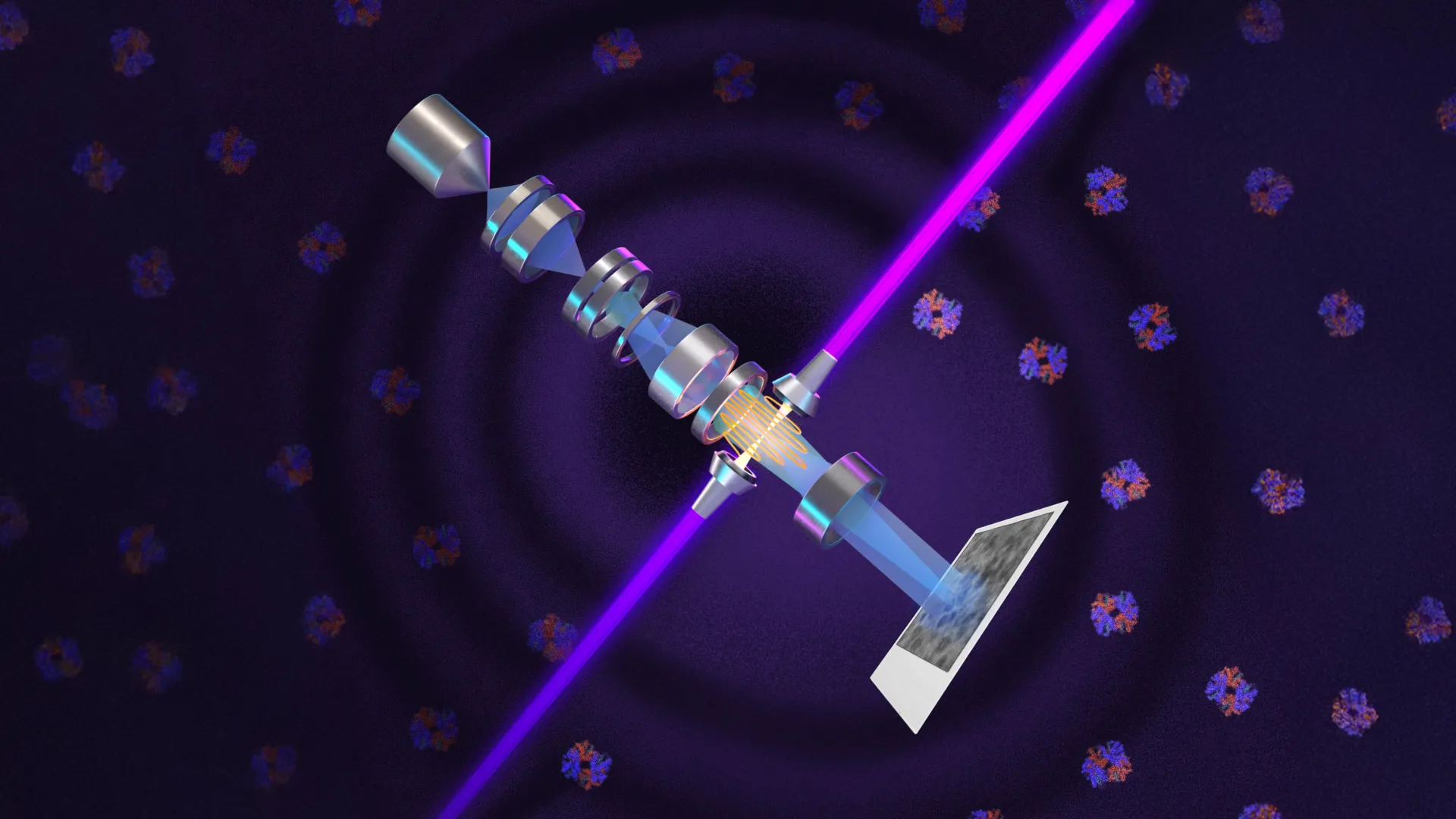
Physicists at the University of California, Berkeley, have announced a significant leap in imaging technology by successfully integrating laser-based phase contrast into electron microscopy. This development, centered on a device known as a laser phase plate, addresses a century-old limitation in biological imaging, potentially allowing scientists to visualize the smallest building blocks of life with unprecedented clarity. By applying the principles that earned the 1953 Nobel Prize in Physics for light microscopy to the much more powerful realm of electron microscopy, the research team has opened a new door into the study of the human proteome and the inner workings of living cells.
The breakthrough, led by Holger Müller, a professor of physics at UC Berkeley and faculty scientist at Lawrence Berkeley National Laboratory, introduces a method to significantly enhance the signal-to-noise ratio in cryoelectron microscopy (cryo-EM). While cryo-EM has already revolutionized structural biology, it has historically struggled to capture clear images of small proteins, which constitute the vast majority of biological molecules in the human body. The new technology, detailed in the journal Science, promises to bridge this gap, potentially enabling the imaging of molecules as small as 17 kilodaltons—a feat previously considered nearly impossible with standard electron optics.
The Evolution of Phase Contrast: From Light to Electrons
To appreciate the magnitude of the Berkeley achievement, one must look back to the early 20th century. In the 1930s, Dutch physicist Frits Zernike observed that biological specimens are often nearly transparent under a microscope because they are composed mostly of water. While light passes through these specimens, it does not significantly dim; however, it does undergo a "phase shift"—a tiny delay in the timing of the light wave’s peaks as it interacts with cellular structures.
Zernike’s genius lay in his ability to convert these invisible phase shifts into visible changes in brightness. By shifting the phase of the unscattered light by 90 degrees, he created high-contrast images of living cells without the need for chemical stains, which typically kill the specimen. This "phase contrast" technique became a cornerstone of biology, earning Zernike the Nobel Prize in 1953.
For decades, scientists sought to replicate this effect in electron microscopes, which use electron beams instead of light to achieve magnifications 10,000 times greater than conventional optical instruments. However, electrons are far more difficult to manipulate than photons. Early attempts to create physical phase plates—often using thin films of carbon—frequently resulted in beam instability, loss of resolution, or the eventual destruction of the plate itself due to the intense radiation of the electron beam.
The solution proposed by Müller and his colleague Robert Glaeser in 2010 was to replace physical hardware with light. They hypothesized that an incredibly intense laser beam could act as a "virtual" phase plate, interacting with the electron beam to induce the necessary 90-degree shift without any physical contact or degradation.
Engineering the World’s Most Intense Continuous Laser Focus
Realizing the 2010 proposal required 15 years of rigorous engineering and a fundamental advancement in laser physics. The primary challenge was generating a laser beam of sufficient intensity to influence an electron beam moving at nearly the speed of light. To achieve the required phase shift, the researchers needed to concentrate an immense amount of energy into a space only a few microns wide.
The Berkeley team’s solution involved a sophisticated optical cavity—a spherical chamber lined with ultra-high-reflectivity mirrors. By bouncing a continuous-wave laser back and forth more than 10,000 times within this cavity, the light intensity builds up to staggering levels. According to Müller, the system generates 75 kilowatts of power focused into a tiny spot. This is significantly more powerful than the lasers used for industrial welding or military applications, creating what is described as the brightest continuous laser focus ever achieved.
This "laser phase plate" is the heart of a new instrument the team has dubbed "Theia," named after the Greek Titaness of light. Built upon a state-of-the-art Thermo Fisher Krios cryo-EM platform, Theia combines advanced electron optics with the laser system to produce a "Formula 1" grade microscope. Unlike previous attempts at phase contrast in electron microscopy, the laser system does not weaken the electron beam or introduce the noise associated with physical apertures.
Breaking the Size Barrier of the Human Proteome
The practical implications of this technology are centered on the "size problem" in structural biology. In the world of molecular biology, mass is measured in daltons. Most current cryo-EM techniques are effective for large protein complexes, but they struggle with molecules smaller than 70 kilodaltons (kDa). This is a critical limitation because approximately 90% of the proteins in the human proteome—the entire set of proteins expressed by our genome—fall below this 70 kDa threshold.
In their published research, the Berkeley team demonstrated the laser phase plate’s efficacy by imaging hemoglobin, a protein responsible for oxygen transport that weighs approximately 64 kDa. While hemoglobin is at the very edge of what conventional cryo-EM can resolve, the laser-enhanced images showed a dramatic improvement in contrast and detail.
"For the most challenging cases—small particles, bad specimens—the laser produces a very considerable advantage," Müller noted. The technology has already pushed the reliable imaging limit down to 50 kDa. The ultimate goal is to reach 17 kDa, the size of myoglobin. Achieving this would effectively "unlock" the vast majority of human proteins for structural analysis, providing pharmaceutical researchers with the blueprints needed to design drugs for diseases that were previously untreatable because their target proteins were "invisible."
Cryo-ET and the Visualization of Cellular Landscapes
While cryo-EM focuses on isolated proteins, the laser phase plate is expected to have an even more profound impact on cryoelectron tomography (cryo-ET). If cryo-EM is like taking a studio portrait of a single person, cryo-ET is like capturing a 3D movie of a crowded city. It involves taking multiple images of a cell from different angles and using computational power to reconstruct a three-dimensional view of how molecules interact in their native environment.
The challenge with cryo-ET is the sheer "crowdedness" of the cell. Bridget Carragher, a leader in the field and founding technical director of imaging at Biohub, compares the task to finding a single leaf on a specific tree within a dense forest. Because the signal-to-noise ratio in these thick cellular samples is so low, it is often impossible to distinguish one protein from the surrounding cellular "noise."
"Cryo-ET needs a dramatic step forward in contrast so we can start to see what’s going on inside the cell," Carragher said. "That’s what the laser phase plate promises to give us."
To further this goal, Biohub is currently constructing a specialized microscope in Redwood City that utilizes a dual-laser design. Building on Müller’s theoretical work, this instrument uses two lasers at right angles to one another, each running at half power. This configuration reduces optical distortions and prevents component burnout, ensuring the microscope can run for long periods—a necessity for the complex data collection required in tomography.
Collaborative Efforts and Industrial Integration
The development of the laser phase plate has been a multi-institutional effort involving UC Berkeley, Lawrence Berkeley National Laboratory, and the Chan Zuckerberg Biohub. Funding from the National Institutes of Health and Biohub was instrumental in moving the project from a theoretical paper in 2010 to a functioning laboratory instrument in 2025.
Crucially, the researchers are working closely with Thermo Fisher Scientific, the world’s leading manufacturer of cryoelectron microscopes. This partnership suggests a clear path for the commercialization and wider distribution of laser phase plate technology. If integrated into standard commercial units, the technology could become a ubiquitous tool in academic and industrial laboratories worldwide.
The Berkeley team’s recent findings, which tested the system against six different biological samples of varying sizes, confirmed that the laser provides a consistent "step function" improvement in image quality. Even for larger proteins where standard cryo-EM is sufficient, the laser phase plate allows for faster data acquisition and higher resolution, making the entire process of structural determination more efficient.
A New Era for Disease Research
The ability to see what was once invisible has historically preceded every major leap in medical science. By providing a 90-degree phase shift to the electron beam, the laser phase plate allows researchers to observe the mechanical operations of molecular machines within living cells for the first time.
Stephani Otte, Biohub’s Vice President of Imaging Science, emphasized the transformative nature of the tool. "We are going to be able to see how molecular machines operate inside the living cell, in context, for the first time," she said. "What was once invisible will become visible—and that changes everything about how we understand disease."
From a diagnostic and therapeutic perspective, this level of detail is invaluable. For instance, researchers could observe how a virus interacts with a cell membrane or how a misfolded protein—the hallmark of Alzheimer’s disease—aggregates within a neuron. By seeing these processes in high resolution and in 3D, scientists can identify new "pockets" on proteins where drugs can bind, potentially leading to more effective treatments with fewer side effects.
As the Berkeley team continues to refine Theia and the Biohub team completes its dual-laser instrument, the focus shifts toward the next frontier: reaching the 17 kDa limit. Müller believes that by further refining the focus of the electron beam and optimizing the laser-electron interaction, the signal-to-noise ratio could be doubled once more. This trajectory suggests that the "golden age" of cryoelectron microscopy is only just beginning, with the laser phase plate serving as the catalyst for a new century of biological discovery.
