
 By Nana O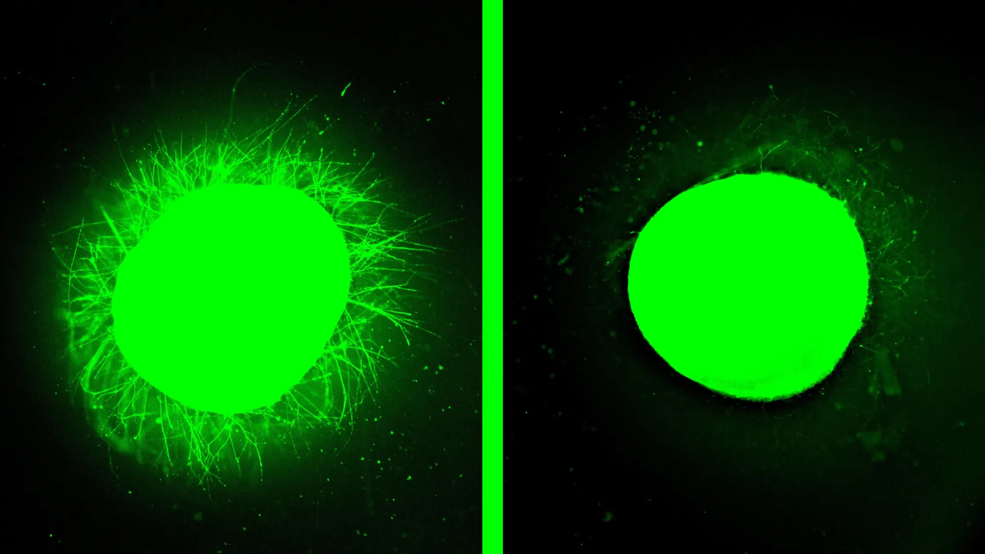
Northwestern University scientists have achieved a significant breakthrough in the quest to understand and treat human spinal cord injuries (SCIs), unveiling the most sophisticated laboratory-grown model of the human spinal cord to date. This innovative organoid system, derived from stem cells, has not only replicated the complex biological devastation of SCI but also demonstrated remarkable therapeutic potential for a novel regenerative treatment. The research, published on February 11 in the prestigious journal Nature Biomedical Engineering, offers a critical new avenue for preclinical drug testing and a renewed sense of hope for individuals living with paralysis.
A Leap Forward in Spinal Cord Injury Research
For decades, understanding the intricate cascade of events following a spinal cord injury has been hampered by the limitations of existing research models. Animal studies, while invaluable, do not fully recapitulate the human biological response. Traditional cell cultures lack the complexity and three-dimensional architecture of the native spinal cord. This new human spinal cord organoid model, however, represents a paradigm shift.
These organoids, measuring several millimeters in diameter, are meticulously engineered from induced pluripotent stem cells (iPSCs). Over months of careful cultivation, researchers guided these stem cells to differentiate into a diverse array of cell types found in the spinal cord, including neurons and astrocytes. Crucially, the team achieved a significant advancement by incorporating microglia, the resident immune cells of the central nervous system, into the organoid structure. This inclusion is vital, as microglia play a pivotal role in the inflammatory response that exacerbates SCI.
"We were the first to introduce microglia into a human spinal cord organoid, so that was a huge accomplishment," stated Samuel I. Stupp, the study’s senior author and a distinguished professor at Northwestern University. "It means that our organoid has all the chemicals that the resident immune system produces in response to an injury. That makes it a more realistic, accurate model of spinal cord injury."
Replicating the Devastation of Injury
The Northwestern team then subjected these advanced organoids to two distinct injury models designed to mimic common forms of human SCI: a scalpel-induced laceration, akin to surgical trauma, and a compressive contusion, mirroring injuries sustained in accidents like car crashes or falls. The results were stark and validated the organoid’s fidelity as a research tool.
Following the simulated trauma, the organoids exhibited key pathological hallmarks of human SCI. Researchers observed significant cell death, a hallmark of acute neuronal damage. A robust inflammatory response was triggered, characterized by the activation of microglia and the release of pro-inflammatory signaling molecules. Perhaps most critically, the organoids developed glial scars.
Glial scarring is a major impediment to functional recovery after SCI. In the injured spinal cord, astrocytes, a type of glial cell, proliferate and form a dense, fibrous barrier. This scar tissue not only physically obstructs regenerating nerve fibers (axons) but also releases inhibitory molecules that actively discourage neuronal regrowth. The Northwestern organoids accurately reproduced this scarring phenomenon, with astrocytes becoming enlarged and densely packed, and the production of chondroitin sulfate proteoglycans (CSPGs), inhibitory molecules associated with nerve injury.
"We could distinguish between the astrocytes that are a part of normal tissue and the astrocytes in the glial scar, which are large and very densely packed," explained Stupp. "We also detected the production of chondroitin sulfate proteoglycans, which are molecules in the nervous system that respond to injury and disease."
This ability to faithfully reproduce the complex cellular and molecular sequelae of SCI in a human-derived model marks a substantial leap beyond previous organoid research, which has primarily focused on basic spinal cord development and disease modeling.
"Dancing Molecules": A Promising Therapeutic Intervention
The true power of the Northwestern model became evident when the researchers applied a novel regenerative therapy known as "dancing molecules" to the injured organoids. This innovative treatment, developed by Stupp and his team, leverages the principles of supramolecular chemistry to stimulate the body’s innate repair mechanisms.
The "dancing molecules" are a type of supramolecular therapeutic peptide (STP). These peptides self-assemble into intricate nanofiber scaffolds that mimic the extracellular matrix of the spinal cord. What sets them apart is their dynamic molecular motion. The molecules are designed to move rapidly and fluidly within this scaffold, even temporarily detaching and reattaching. This dynamic movement is crucial for effectively interacting with cell receptors, which are themselves constantly in motion.
"Given that cells themselves and their receptors are in constant motion, you can imagine that molecules moving more rapidly would encounter these receptors more often," Stupp had previously stated, referring to earlier animal studies. "If the molecules are sluggish and not as ‘social,’ they may never come into contact with the cells."
In the current study, when the injured spinal cord organoids were treated with these "dancing molecules," the results were nothing short of dramatic. The dense glial scar tissue significantly diminished, becoming barely detectable. Simultaneously, the organoids exhibited substantial neurite outgrowth – the regeneration of axons, the long, thread-like extensions that enable neurons to communicate with each other. These regenerated neurites began to grow in organized patterns, suggesting a potential for functional reconnection.
This finding strongly supports the therapeutic potential of "dancing molecules." In previous animal studies, a single injection of this therapy administered 24 hours after a severe SCI in mice led to significant motor recovery, with the animals regaining the ability to walk within four weeks. Formulations exhibiting faster molecular motion consistently outperformed those with slower movement, underscoring the critical role of dynamic molecular interactions in driving bioactivity and cellular signaling.
The "dancing molecules" therapy recently received Orphan Drug Designation from the U.S. Food and Drug Administration (FDA), a designation that provides incentives and support for the development of drugs for rare diseases or conditions, underscoring the significant therapeutic promise it holds.
The Significance of Human Organoids in Drug Development
The implications of this research extend far beyond the immediate findings. The ability to reliably test new therapies in a human spinal cord model in vitro (in a lab dish) before advancing to human clinical trials is a monumental step forward.
"One of the most exciting aspects of organoids is that we can use them to test new therapies in human tissue," emphasized Stupp. "Short of a clinical trial, it’s the only way you can achieve this objective. We decided to develop two different injury models in a human spinal cord organoid and test our therapy to see if the results resembled what we previously saw in the animal model. After applying our therapy, the glial scar faded significantly to become barely detectable, and we saw neurites growing, resembling the axon regeneration we saw in animals. This is validation that our therapy has a good chance of working in humans."
Organoids offer several advantages over traditional research methods. They are derived from human stem cells, providing a more accurate representation of human biology than animal models. Their development and testing are also generally more cost-effective and can be conducted at a faster pace than animal experiments or human clinical trials. This allows researchers to rapidly screen potential therapeutic candidates, identify promising leads, and optimize treatment strategies.
A Look Towards the Future: Personalized Medicine and Chronic Injuries
The Northwestern team is not resting on its laurels. They have ambitious plans to further refine and expand their organoid model. Future research will focus on engineering even more complex organoids capable of replicating chronic, long-standing spinal cord injuries. These injuries typically involve more extensive and recalcitrant scar tissue, presenting a greater therapeutic challenge.
Furthermore, the long-term vision includes the potential for personalized medicine. By generating organoids from a patient’s own stem cells, researchers could develop and test therapies tailored to an individual’s specific genetic makeup and injury profile. This approach could significantly reduce the risk of immune rejection, a common complication in transplantation therapies.
"With further development, these miniature spinal cords could contribute to personalized medicine by generating implantable tissue from a patient’s own stem cells, reducing the risk of immune rejection," Stupp projected.
The study was supported by the Center for Regenerative Nanomedicine at Northwestern University and a generous gift from the John Potocsnak Family for spinal cord injury research, highlighting the critical role of dedicated funding in advancing such groundbreaking scientific endeavors.
This pioneering work at Northwestern University represents a beacon of hope in the challenging landscape of spinal cord injury research, offering a tangible path towards developing effective treatments that could one day restore function and improve the lives of millions worldwide. The combination of a highly sophisticated human organoid model and a dynamically active regenerative therapy marks a significant turning point, moving the field closer to translating laboratory discoveries into clinical realities.
