
 By George Ongkojoyo
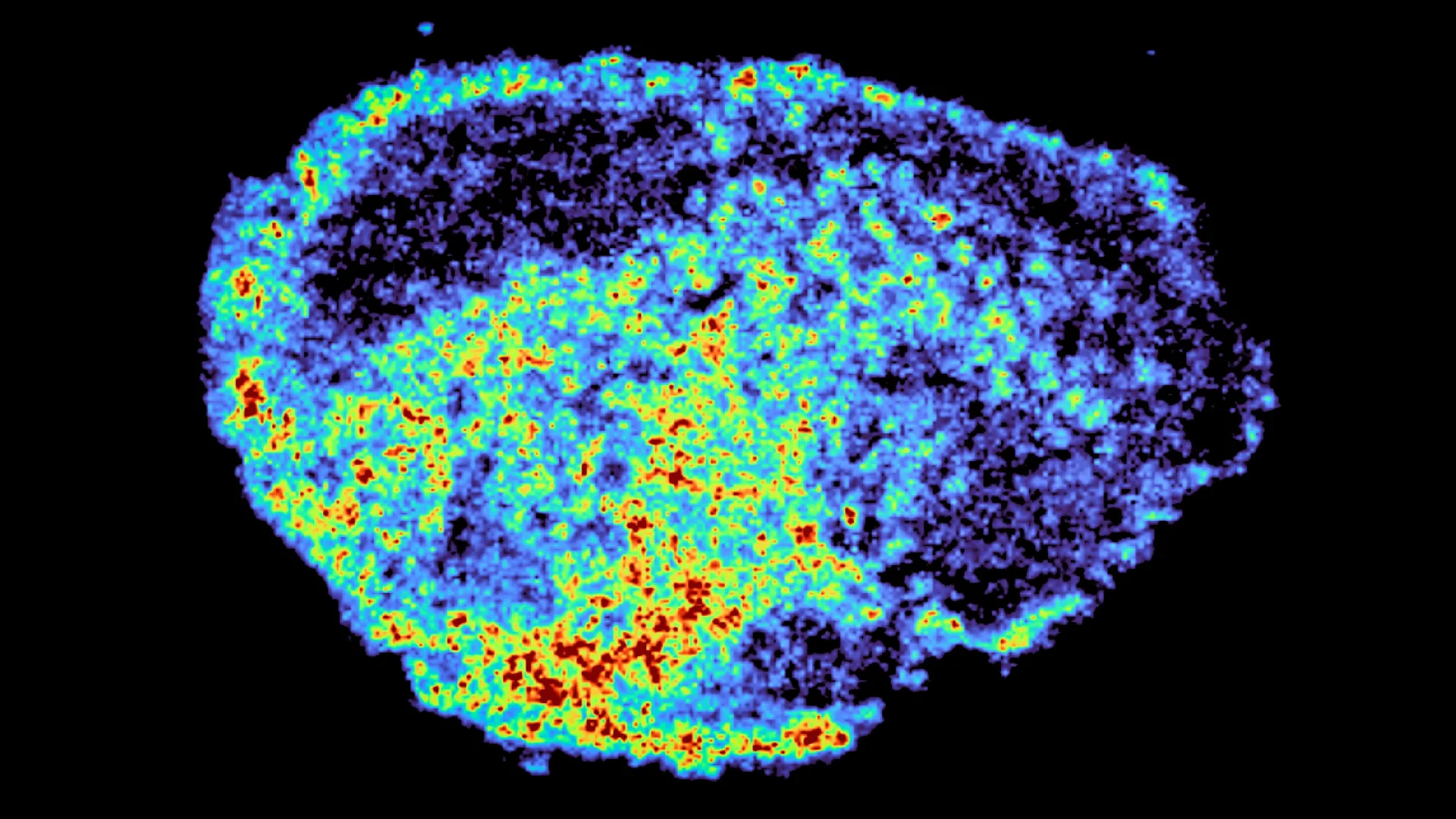
A landmark study published in the journal Nature Communications has unveiled a critical mechanism explaining why some cancer patients fail to respond to targeted therapies even when the drugs successfully reach the tumor site. Led by Dr. Louise Fets at the MRC Laboratory of Medical Sciences (LMS), the research identifies the role of lysosomes—small, acidic organelles within cells—as internal reservoirs that can trap and slowly release specific cancer drugs. This discovery, centered on a class of drugs known as PARP inhibitors, provides a new framework for understanding drug resistance and the uneven distribution of medicine within human tissue. By utilizing advanced imaging techniques such as mass spectrometry imaging and spatial transcriptomics, the research team demonstrated that the effectiveness of a treatment is dictated not just by its presence in the tumor, but by its precise sub-cellular location and the biological "pockets" it occupies.
The Evolution and Mechanism of PARP Inhibitors in Oncology
To understand the significance of the findings, it is essential to contextualize the role of PARP (poly ADP-ribose polymerase) inhibitors in modern oncology. PARP inhibitors represent one of the most successful applications of the concept of "synthetic lethality." These drugs work by blocking the PARP enzyme, which is responsible for repairing single-strand breaks in DNA. In patients with specific genetic mutations, such as those in the BRCA1 or BRCA2 genes, the cell’s alternative DNA repair mechanism (homologous recombination) is already defective. When a PARP inhibitor is introduced, the cell becomes unable to repair any DNA damage, leading to genomic instability and eventual programmed cell death.
Since the first PARP inhibitor, olaparib, received FDA approval in 2014, the therapeutic landscape for ovarian, breast, prostate, and pancreatic cancers has been transformed. Subsequent approvals of rucaparib and niraparib expanded the options for clinicians. However, clinical data has consistently shown a spectrum of responses. While some patients experience long-term remission, others see their tumors continue to grow, or they develop resistance after an initial period of success. Until now, much of the research into this resistance has focused on genetic mutations that allow cancer cells to bypass the PARP blockade. The new study from the MRC LMS suggests that the physical distribution of the drug itself—pharmacokinetics at a microscopic level—may be equally influential.
Chronology of the Research and Methodology
The research project was conceived to address a fundamental gap in pharmacological knowledge: the "black box" of drug behavior once it crosses the cell membrane in a complex tumor environment. Traditional methods of measuring drug concentration often rely on "bulk" analysis, where a piece of tissue is ground up and the average amount of drug is measured. This method, however, masks the extreme variations that can occur between neighboring cells.
The study followed a multi-stage chronological progression:
- Explant Preparation: The team utilized fresh ovarian tumor samples obtained from patients. Rather than using simplified 2D cell cultures, they employed "explants"—thin slices of live tumor tissue. This allowed the researchers to maintain the structural integrity and cellular diversity of the original cancer environment.
- Drug Treatment and Incubation: These explants were treated with clinically relevant doses of three primary PARP inhibitors: olaparib, rucaparib, and niraparib.
- Mass Spectrometry Imaging (MSI): After treatment, the samples were analyzed using MSI. This high-resolution technique allows scientists to visualize the exact spatial coordinates of drug molecules across the tissue slice.
- Spatial Transcriptomics Integration: In a sophisticated technological pairing, the researchers matched the drug maps with spatial transcriptomics. This allowed them to see which genes were being expressed in areas where the drug was highly concentrated versus areas where it was sparse.
- Sub-cellular Analysis: Finally, the team focused on the organelles within the cells to determine why certain drugs were clustering in specific zones.
Data Analysis: The Lysosomal Reservoir Effect
The core finding of the study revolves around the lysosome, often referred to as the cell’s "recycling center" or "garbage disposal." These organelles are highly acidic environments designed to break down waste products. The researchers discovered that certain PARP inhibitors are "lysosmotropic," meaning they are chemically drawn to and trapped within these acidic compartments.
The data revealed a striking divergence in how different PARP inhibitors behave:
- Rucaparib and Niraparib: These drugs showed a high affinity for lysosomes. In the tumor samples, they did not distribute evenly. Instead, they accumulated in high concentrations within these cellular pockets. The researchers noted that these lysosomes acted as "slow-release reservoirs," holding the drug and then leaking it out over time.
- Olaparib: In contrast, olaparib did not show the same level of lysosomal sequestration. It distributed more uniformly across the tissue, suggesting that its chemical structure makes it less susceptible to being trapped by the cell’s internal acidity.
This uneven distribution creates a "patchwork" effect within the tumor. Some cells are saturated with the drug, potentially leading to higher toxicity or enhanced efficacy, while neighboring cells receive sub-therapeutic doses, allowing them to survive and potentially mutate into drug-resistant strains. The study quantified this variability, noting that drug levels could vary significantly even between cells that were only micrometers apart.
Official Responses and Expert Insights
The implications of the study have drawn significant attention from the academic and medical communities. Dr. Zoe Hall, an Associate Professor at Imperial College London’s Department of Metabolism, Digestion and Reproduction and a senior author of the study, emphasized the technological breakthrough of the research. "A novel aspect of this study was the use of mass spectrometry imaging to directly measure and visualize drug uptake in patient tumour tissue," Dr. Hall stated. She noted that the ability to compare gene expression and drug mapping on the same tissue slice provides a level of detail previously unattainable in clinical oncology.
Dr. Carmen Ramirez Moncayo, the study’s first author, highlighted the unexpected nature of the findings regarding single-cell variability. "We were surprised to see large variability in drug accumulation at the single-cell level," she said. "This variability was driven by the build-up of a drug in lysosomes, which are acting as reservoirs, increasing the exposure of cancer cells to drugs, by storing and releasing the drug when needed."
Dr. Louise Fets, Head of the Drug Transport and Tumour Metabolism Group at the LMS, framed the research within the broader goal of personalized medicine. She expressed hope that by understanding the "molecular signature" of a patient’s tumor—including its lysosomal activity—doctors could eventually tailor the choice of PARP inhibitor to the specific biological profile of the individual.
Broader Impact on Future Cancer Therapeutics
The discovery that lysosomes act as drug reservoirs has several far-reaching implications for the future of cancer care and drug development.
1. Optimization of Drug Choice:
The fact that different PARP inhibitors interact differently with lysosomes suggests that they are not interchangeable. If a patient’s tumor is characterized by a high density of lysosomes or specific acidic profiles, a non-lysosmotropic drug like olaparib might be more effective in ensuring even distribution. Conversely, in cases where a "sustained release" effect is desired, rucaparib or niraparib might offer advantages.
2. Overcoming Resistance:
One of the primary drivers of cancer relapse is the survival of a small population of "persister" cells. If these cells survive because they are located in "cold spots" where drug distribution is low due to lysosomal trapping in neighboring cells, then strategies to modify lysosomal pH or inhibit sequestration could potentially sensitize the entire tumor to treatment.
3. Enhancing Drug Design:
Pharmaceutical companies can use this data to refine the chemical structures of next-generation inhibitors. By adjusting the basicity or lipophilicity of a molecule, chemists can intentionally design drugs to either seek out or avoid lysosomal accumulation, depending on the therapeutic goal.
4. Advancing Spatial Biology:
This study reinforces the growing importance of "spatial biology"—the study of how the location of cells and molecules influences health and disease. As oncology moves away from bulk tissue analysis toward high-resolution spatial mapping, the ability to predict drug behavior in a complex 3D environment will become a standard part of drug testing.
Limitations and Next Steps
While the study provides a major leap forward, the researchers acknowledged the limitations of working with "explant" models. In a living patient, the delivery of drugs is governed by the circulatory system. Cancerous tumors are notorious for having "leaky" and disorganized blood vessels, which creates further barriers to even drug distribution. The next phase of research will involve moving into animal models to observe how blood flow and the tumor microenvironment interact with the lysosomal trapping mechanism in real-time.
Furthermore, the team plans to expand the study to include larger cohorts of patients, particularly those who have already relapsed. By comparing the drug-mapping profiles of primary tumors versus relapsed tumors, they hope to determine if changes in lysosomal behavior are a primary driver of acquired resistance.
The research was a collaborative effort supported by a diverse range of institutions, including the Medical Research Council, Cancer Research UK, and the Victoria’s Secret Global Fund for Women’s Cancers. As the medical community continues to strive for more effective and less toxic treatments, the insights provided by Dr. Fets and her team offer a vital new map for navigating the complex interior of the cancer cell. Through the lens of spatial transcriptomics and mass spectrometry, the "recycling centers" of the cell have now been revealed as key players in the fight against ovarian cancer, potentially changing the way targeted therapies are administered and monitored in the years to come.
