
 By Gabriel Paijo
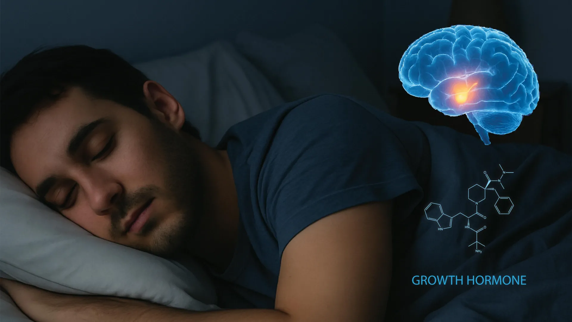
The intricate relationship between the human brain, the quality of restorative sleep, and the endocrine system has long been a subject of intense scientific inquiry. For decades, clinicians have observed that a lack of sleep leads to stunted growth in children and metabolic dysfunction in adults, yet the specific neurological "wiring" that connects these two systems remained elusive. A landmark study led by researchers at the University of California, Berkeley, and published in the prestigious journal Cell, has finally mapped the neural circuitry responsible for growth hormone (GH) release during sleep. This discovery not only clarifies how the brain triggers hormone production during the deepest stages of rest but also reveals a surprising feedback loop where growth hormone itself acts as a regulator for wakefulness.
By identifying the specific neurons within the hypothalamus and their interaction with the brainstem, the Berkeley team has provided a blueprint that could revolutionize the treatment of sleep disorders, metabolic diseases, and neurodegenerative conditions. The findings underscore the biological reality that sleep is not a passive state of inactivity, but a highly coordinated physiological process essential for physical repair and chemical balance.
The Biological Significance of Growth Hormone and Deep Sleep
Growth hormone, secreted by the pituitary gland, is a cornerstone of human physiology. While its most visible role is during puberty—where it facilitates the lengthening of bones and the development of muscle tissue—its utility persists throughout the human lifespan. In adults, GH is responsible for regulating fat metabolism, maintaining bone density, and ensuring the body effectively manages glucose levels.
Historically, the medical community has recognized that GH secretion is pulsatile, with the largest and most significant pulse occurring shortly after the onset of deep, non-rapid eye movement (non-REM) sleep. This stage, often referred to as slow-wave sleep (SWS), is characterized by high-amplitude brain waves and is the period when the body performs its most intensive "maintenance" work. When this sleep stage is truncated or fragmented—common in modern society due to artificial lighting, stress, and sleep apnea—growth hormone levels plummet. This deficiency is a known precursor to increased visceral fat, reduced muscle mass, and a higher risk of cardiovascular disease.
The UC Berkeley study sought to move beyond the "what" of this relationship to the "how." By focusing on the hypothalamus, a pea-sized region at the base of the brain that acts as the command center for the autonomic nervous system, researchers identified the precise switches that turn GH production on and off.
Mapping the Hypothalamic Circuitry
The research, conducted in the laboratory of Yang Dan, a professor of molecular and cell biology and a Howard Hughes Medical Institute investigator, focused on two primary types of neurons within the hypothalamus: those that produce growth hormone-releasing hormone (GHRH) and those that produce somatostatin.
In a healthy system, these two messengers exist in a state of push-and-pull. GHRH acts as the "accelerator," signaling the pituitary gland to release GH into the bloodstream. Somatostatin acts as the "brake," inhibiting that release. The Berkeley team discovered that during the transition into deep sleep, the brain orchestrates a specific dance between these neurons.
During non-REM sleep, somatostatin activity decreases significantly, effectively "lifting the brake." Simultaneously, GHRH activity rises, providing the necessary signal for a surge of growth hormone. Interestingly, the researchers found that during REM sleep—the stage associated with dreaming—both GHRH and somatostatin activity actually increase. While they are opposing forces, their high-level co-activation during REM still contributes to the overall hormonal output, though through a different mechanical pathway than seen in non-REM sleep.
The Discovery of a Bidirectional Feedback Loop
The most groundbreaking aspect of the study is the discovery that growth hormone does not merely take orders from the brain; it talks back. The researchers identified a previously unknown feedback system involving the locus coeruleus (LC), a cluster of neurons in the brainstem.
The locus coeruleus is the brain’s primary source of norepinephrine, the chemical responsible for arousal, alertness, and the "fight or flight" response. It is the region that tells the brain when it is time to wake up and engage with the environment. The Berkeley study revealed that as growth hormone levels rise during a period of deep sleep, the hormone eventually begins to stimulate the locus coeruleus.
"Sleep drives growth hormone release, and growth hormone feeds back to regulate wakefulness," explained Daniel Silverman, a UC Berkeley postdoctoral fellow and study co-author. This creates a homeostatic balance. If the body has received enough deep sleep to facilitate the necessary growth and repair, the resulting rise in GH acts as a chemical signal to the brain that it is time to transition toward wakefulness.
This finding suggests that growth hormone acts as a "sleep-o-meter," helping the brain gauge the adequacy of its rest. It provides a biological explanation for why people often feel a natural "surge" of alertness after a night of high-quality, uninterrupted sleep, compared to the grogginess associated with sleep that is long in duration but poor in hormonal quality.
Methodology: Using Optogenetics to Trace Neural Pathways
To reach these conclusions, the Berkeley team utilized advanced neuroscientific techniques, including optogenetics and in-vivo electrode recording in mice. Mice serve as an excellent model for this research because the fundamental architecture of the hypothalamus and the locus coeruleus has been conserved across mammalian evolution.
The researchers implanted micro-electrodes to record neural activity in real-time while the mice moved through their natural sleep-wake cycles. Because mice sleep in short bursts rather than one consolidated block, the team was able to observe hundreds of transitions between sleep stages and waking states. Using light-sensitive proteins (optogenetics), the scientists could "turn on" or "turn off" specific GHRH or somatostatin neurons with laser precision.
This level of control allowed them to prove causality. When they artificially stimulated the GHRH neurons, they observed a direct increase in GH and a subsequent influence on the locus coeruleus. Conversely, by manipulating the locus coeruleus, they could see how the brain’s arousal center responded to the hormonal signals.
Chronology of Scientific Understanding
The Berkeley study represents the culmination of decades of incremental progress in the fields of endocrinology and neuroscience:
- 1960s: Researchers first identify the correlation between slow-wave sleep and growth hormone peaks in humans using early EEG technology and blood sampling.
- 1980s: The chemical structure of GHRH is first isolated, allowing scientists to understand the pituitary gland’s triggers.
- 2000s: The rise of optogenetics allows for the mapping of specific brain circuits with millisecond precision, moving beyond general observations of brain regions.
- Current Study: The UC Berkeley team bridges the gap between the hypothalamus (hormone control) and the locus coeruleus (arousal control), identifying the bidirectional feedback loop for the first time.
Implications for Metabolic and Neurological Health
The implications of this research extend far beyond basic biology. By understanding the "handle" that controls growth hormone release and the locus coeruleus, scientists may develop new ways to treat a variety of debilitating conditions.
1. Metabolic Disorders and Diabetes
Since growth hormone is a critical regulator of glucose and fat metabolism, chronic sleep disruption—which leads to GH suppression—is a major contributor to the global obesity and diabetes epidemics. "Inadequate sleep may contribute to an increased risk of obesity and cardiovascular disease because the hormone helps control how the body uses stored fat," the study notes. Future therapies could potentially involve targeting these specific hypothalamic neurons to restore hormonal balance in patients with metabolic syndrome.
2. Neurodegenerative Diseases
The locus coeruleus is one of the first areas of the brain to show signs of degeneration in patients with Alzheimer’s and Parkinson’s diseases. Because this study identifies a link between GH and LC activity, it opens the door to investigating whether maintaining healthy growth hormone levels could protect the locus coeruleus from early-onset decay.
3. Sleep Disorders
For individuals suffering from insomnia or fragmented sleep, the discovery offers a new target for pharmaceutical intervention. Instead of general sedatives that often bypass natural sleep architecture, new treatments could focus on "dialing back" the excitability of the locus coeruleus or modulating GHRH to encourage more natural, restorative sleep cycles.
Analysis: The Delicate Balance of Homeostasis
The research highlights the concept of homeostasis—the body’s innate drive to maintain a stable internal environment. The discovery that "too much growth hormone can push the brain toward wakefulness" provides a cautionary tale for the off-label use of growth hormone supplements. If the balance is artificially tipped, it could lead to chronic over-arousal and insomnia, creating a vicious cycle where the body cannot reach the deep sleep stages required to process the very hormones being supplemented.
Furthermore, the study sheds light on the "paradoxical" nature of the locus coeruleus. While it typically promotes wakefulness, earlier research by Daniel Silverman showed that over-excitation of this region can sometimes cause the brain to "shut down" into sleep as a protective mechanism. This complexity suggests that any future clinical treatments must be finely tuned to the individual’s existing neural landscape.
Conclusion and Future Directions
The work of Xinlu Ding, Daniel Silverman, Yang Dan, and their colleagues at UC Berkeley and Stanford University marks a significant shift in our understanding of the sleeping brain. By moving from blood-level observations to direct neural recordings, they have revealed that the brain and the endocrine system are in a constant, high-speed dialogue.
"Growth hormone not only helps you build your muscle and bones and reduce your fat tissue, but may also have cognitive benefits, promoting your overall arousal level when you wake up," said first author Xinlu Ding. This research reaffirms that the quality of our sleep is a fundamental pillar of our physical and mental health.
As the scientific community moves forward, the next step will be to translate these findings from mouse models to human clinical trials. While the underlying circuitry is similar, the complexities of human lifestyle, aging, and disease will require further exploration. For now, the discovery serves as a powerful reminder that the body’s most important work—growing, repairing, and balancing—happens while we are most vulnerable, lost in the depths of a good night’s sleep.
