
 By Budi Oetomo Narendra
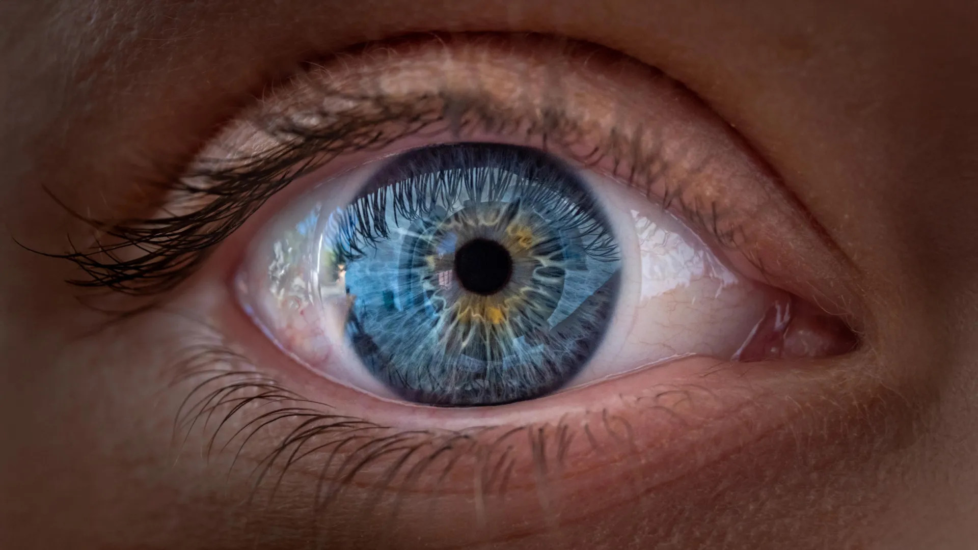
A landmark discovery by scientists at Johns Hopkins University has fundamentally reshaped our understanding of how the intricate architecture of human vision develops. Their groundbreaking research, published in the Proceedings of the National Academy of Sciences, reveals a previously unappreciated and surprising role for a vitamin A derivative, retinoic acid, working in concert with thyroid hormones, in the formation of the foveola – the critical part of the eye responsible for our sharpest, most detailed sight. This revelation directly challenges a long-standing scientific explanation that has held sway for approximately 30 years, opening new avenues for both basic research into ocular development and potential therapeutic strategies for debilitating vision loss conditions.
Unraveling the Mystery of Central Vision Development
The core of this discovery lies in an unexpected cellular transformation occurring within the developing retina before birth. Specifically, certain light-detecting cells, initially configured to sense blue light, appear to undergo a remarkable metamorphosis, converting into cells sensitive to red and green light. This intricate cellular reprogramming is crucial for establishing the specialized arrangement of photoreceptors necessary for the exceptionally clear and detailed central vision that defines human sight.
The findings are poised to significantly alter scientific paradigms regarding human eye development and could accelerate research into future treatments for prevalent conditions such as macular degeneration, glaucoma, and other diseases that severely impair vision. Dr. Robert J. Johnston Jr., an associate professor of biology at Johns Hopkins University and the lead author of the study, emphasized the profound implications of this work. "This is a key step toward understanding the inner workings of the center of the retina, a critical part of the eye and the first to fail in people with macular degeneration," Dr. Johnston stated. "By better understanding this region and developing organoids that mimic its function, we hope to one day grow and transplant these tissues to restore vision."
The Foveola: A Microscopic Marvel of Vision
To appreciate the significance of this discovery, it is essential to understand the foveola. Located at the very center of the macula, which itself is part of the retina, the foveola is a tiny, pit-like depression. The retina, a thin layer of tissue at the back of the eye, acts like the film in a camera, converting light into electrical signals that are sent to the brain. Within this complex structure, the foveola stands out for its unparalleled role in high-acuity central vision. It enables humans to perform tasks requiring fine detail discrimination, such as reading, recognizing faces, and appreciating intricate visual information.
Despite occupying only a minuscule fraction of the entire retina, the foveola is disproportionately vital, contributing to approximately 50% of overall human visual perception. This remarkable efficiency is due to its unique cellular composition and architecture. The retina contains two main types of photoreceptor cells: rods and cones. Rods are highly sensitive to light and are crucial for vision in dim conditions, while cones are responsible for daytime vision and the perception of color.
Humans possess three types of cone cells, each optimally sensitive to different wavelengths of light, colloquially known as blue, green, and red cones. The brain interprets signals from these different cone types to construct a vast spectrum of colors. However, these cones are not uniformly distributed. The foveola is uniquely characterized by a high density of red and green cones, but it conspicuously lacks blue cones, which are relegated to the peripheral regions of the retina. This precise, asymmetric distribution is the biological foundation for the foveola’s exceptional sharpness and clarity of vision.
A Longstanding Enigma and the Limitations of Traditional Research
For many decades, the scientific community has grappled with explaining how this highly specialized and asymmetric pattern of cone distribution in the foveola develops. A significant hurdle in this quest has been the lack of suitable animal models. Commonly studied animals, including rodents and fish, do not share the same foveolar organization as humans, making it exceedingly difficult to extrapolate findings from animal experiments to human ocular development. This limitation has left a critical gap in our understanding of how our most sophisticated visual capabilities are forged.
The prevailing hypothesis for roughly 30 years posited that blue cones, which might initially appear in the developing foveola, would simply migrate away as the retina matured. This model was based on the assumption that once a cone cell committed to a particular identity (e.g., blue-sensitive), that identity remained fixed and immutable throughout development. The new research, however, paints a much more dynamic and surprising picture.
Innovative Organoid Technology: A Window into Human Development
To overcome the challenges posed by traditional animal models and peer into the intricate processes of human retinal development, Dr. Johnston and his team employed an innovative laboratory technique they have been instrumental in developing over several years: the cultivation of miniature, self-organizing human retinal tissues known as organoids.
These small, three-dimensional clusters of cells, derived from human fetal cells, possess the remarkable ability to spontaneously reproduce many of the key structural and functional features of developing human tissue. By meticulously growing and observing these retinal organoids over several months, the scientists gained an unprecedented opportunity to track complex cellular changes that would otherwise be practically impossible to study directly within a developing human fetus during pregnancy. This cutting-edge methodology provided a crucial window into the precise chronological events governing foveolar formation.
The Chronology of Transformation: Weeks 10-14 of Fetal Development
The meticulous observation of these retinal organoids revealed a fascinating developmental chronology. During weeks 10 through 12 of fetal development, a small number of blue cones are indeed initially present within the region destined to become the foveola. However, by week 14, a dramatic shift occurs: these blue-sensitive cells appear to have undergone a remarkable transformation, changing their identity to become red and green cones. This observation directly contradicted the long-held migration theory and hinted at a more complex, plastic cellular process.
The team then delved into identifying the molecular orchestrators behind this profound cellular identity change. Their investigation pinpointed two critical chemical influences acting in a coordinated manner.
The Dual Role of Retinoic Acid and Thyroid Hormones
The first key player identified is retinoic acid, a powerful signaling molecule derived from vitamin A. Retinoic acid is well-known for its fundamental roles in regulating cell growth, differentiation, and specialization across various tissues throughout the body. In the context of the developing retina, the researchers discovered that the precise breakdown and metabolism of retinoic acid in the central region actively limits the initial formation of blue cones there. This acts as an early patterning mechanism, setting the stage for the specialized foveolar composition.
The second crucial influence comes from thyroid hormones. While primarily recognized for their roles in regulating metabolism, growth, and overall development, thyroid hormones also exert specific effects on cellular differentiation. In this particular instance, the hormones appear to actively encourage and drive the remaining blue cones within the foveola to undergo their identity switch, transforming into red and green cones.
Dr. Johnston succinctly summarized this elegant two-step process: "First, retinoic acid helps set the pattern. Then, thyroid hormone plays a role in converting the leftover cells." He further emphasized the functional significance, stating, "That’s very important because if you have those blue cones in there, you don’t see as well."
It is important to clarify that this discovery pertains to the intricate processes of eye development before birth. The findings do not imply that taking vitamin A supplements can enhance or improve sharp vision in adults, as the developmental windows and mechanisms are distinct.
A Direct Challenge to a 30-Year Paradigm
The implications of these results are profound, offering a striking alternative to a scientific explanation that has shaped vision research for decades. The longstanding model, established approximately 30 years ago, proposed that any blue cones initially present in the foveola simply migrated out of the region as the retina matured. This model was predicated on the assumption that once a photoreceptor cell committed to a specific type of cone, its identity remained fixed and unchangeable throughout its lifespan.
The new findings, however, present compelling evidence for a different, more dynamic reality. Rather than merely relocating, at least some blue cones in the developing foveola appear to undergo a genuine change in identity, converting into red or green cones. This suggests a remarkable degree of cellular plasticity previously unrecognized in the context of photoreceptor development.
"The main model in the field from about 30 years ago was that somehow the few blue cones you get in that region just move out of the way, that these cells decide what they’re going to be, and they remain this type of cell forever," Dr. Johnston explained. "We can’t really rule that out yet, but our data supports a different model. These cells actually convert over time, which is really surprising." While the researchers acknowledge that some cellular migration cannot be entirely ruled out, their observations strongly support the notion that developing cone cells possess a far greater degree of flexibility and adaptability than previously understood. This paradigm shift is poised to fundamentally alter how scientists approach the study of human color vision formation and the development of the retina’s most specialized and critical regions.
Broader Impact: Towards Restoring Lost Vision
Beyond providing a clearer mechanistic explanation for healthy eye development, these findings carry significant implications for the treatment of diseases that devastate the retina and cause vision loss.
One particularly promising application lies in the battle against macular degeneration, a leading cause of central vision loss, especially among older adults. This progressive disease damages the macula, the very retinal region responsible for detailed central vision, profoundly interfering with daily activities such as reading, driving, and recognizing faces. While certain treatments can slow or manage specific forms of the disease, a definitive cure remains elusive. Understanding the precise molecular and cellular mechanisms by which the retina creates and arranges its specialized light-sensitive cells could eventually pave the way for laboratory-grown, healthy replacements.
Dr. Johnston’s team is actively engaged in refining their retinal organoid models, striving to make them even more closely mimic the intricate structure and complex functions of the human eye. The long-term vision is to develop precisely controlled populations of photoreceptors – the light-detecting cells – suitable for potential transplantation into damaged retinas.
Katarzyna Hussey, a study author and former doctoral student in Dr. Johnston’s laboratory, now a molecular and cell biologist at CiRC Biosciences, a cell therapy company in Chicago, expressed optimism about this novel direction for vision restoration. "The goal with using this organoid tech is to eventually make an almost made-to-order population of photoreceptors," Hussey noted. "A big avenue of potential is cell replacement therapy to introduce healthy cells that can reintegrate into the eye and potentially restore that lost vision."
The premise of cell replacement therapy involves introducing healthy, lab-grown photoreceptor cells into an eye damaged by disease, with the hope that these new cells can integrate with the existing retinal tissue, establish proper connections with the eye’s neural circuitry, and safely restore lost functions. This approach represents a monumental scientific and medical endeavor.
Hussey, while enthusiastic about the potential, underscored the substantial development and rigorous testing still required before such technology could reach routine clinical use. "These are very long-term experiments, and of course we’d need to do optimizations for safety and efficacy studies prior to moving into the clinic. But it’s a viable journey."
The scientific community is likely to greet these findings with considerable excitement. The ability to precisely control the differentiation and patterning of human photoreceptors in vitro offers an unprecedented tool for drug screening, disease modeling, and ultimately, regenerative medicine. This discovery could ignite renewed interest and funding in the study of retinal development, drawing in researchers from diverse fields such as developmental biology, ophthalmology, genetics, and bioengineering. The detailed mechanistic insights provided by Johnston’s team could accelerate the timeline for developing targeted therapies that not only slow disease progression but actively restore lost visual function.
For now, this fundamental discovery provides something invaluable: a significantly clearer and more nuanced explanation of how the human eye develops its remarkable capacity for seeing fine details. By meticulously uncovering how vitamin A signaling and thyroid hormones orchestrate the intricate cellular ballet within the developing retina, scientists at Johns Hopkins have taken a critical stride forward in understanding vision at its earliest stages, bringing humanity closer to potentially finding ways to preserve and restore this most precious sense when disease inevitably strikes.
