
 By Nana O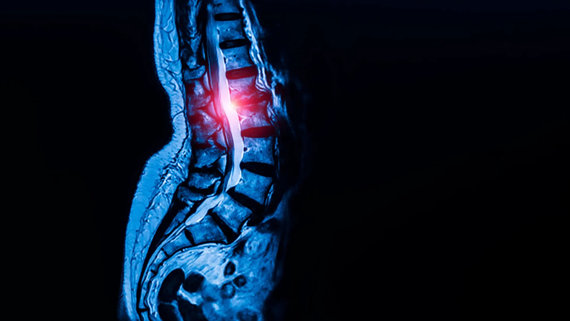
Researchers at Weill Cornell Medicine and the Hospital for Special Surgery (HSS) have pinpointed a previously elusive population of stem cells that are instrumental in the development and maintenance of the body’s tendons and ligaments. This groundbreaking discovery, published on September 7 in the prestigious journal Cell, not only sheds light on the fundamental biology of connective tissues but also offers a promising new therapeutic target for a debilitating condition: lumbar spinal stenosis. The findings suggest that an overactivity of these specialized stem cells in the lower spine may be a key driver of this widespread ailment, potentially paving the way for novel drug-based treatments.
Lumbar spinal stenosis is a significant global health concern, affecting an estimated 103 million people worldwide. The condition arises when the space within the spinal canal narrows, often due to the thickening of ligaments. This constriction can impinge upon spinal nerves, leading to a cascade of symptoms including chronic pain, numbness, and profound difficulty with mobility, severely impacting quality of life. Currently, surgical intervention is often the primary recourse for patients with advanced stenosis, a prospect that carries inherent risks and recovery burdens.
The scientific journey leading to this discovery involved years of meticulous research, building upon previous investigations into skeletal stem cells. Dr. Matthew Greenblatt, co-corresponding author and the Rohr Family Research Scholar, associate professor of pathology and laboratory medicine at Weill Cornell and a pathologist at NewYork-Presbyterian/Weill Cornell Medical Center, explained the challenges in identifying these specific cells. "While previous studies had proposed several candidate stem cells, none had definitively shown that a single cell population could both self-renew and generate the full spectrum of tendon and ligament cell types," Dr. Greenblatt stated. This elusive nature stemmed from the inherent similarity in appearance of many fibroblast-like cells within these connective tissues, making differentiation and isolation a formidable task.
Unraveling the Mystery of Connective Tissue Stem Cells
The research team employed advanced single-cell analysis techniques to overcome these challenges. By meticulously analyzing thousands of individual cells and sorting them into distinct cell types, they were able to identify a specific population exhibiting the hallmarks of "stemness." This crucial characteristic denotes the cell’s capacity for continuous self-renewal while simultaneously producing the diverse array of mature cells required for the formation and ongoing repair of tendons and ligaments.
Initial investigations in mouse models revealed these specialized stem cells residing in discrete, specialized niches within tendons and ligaments. These niches appear to function as crucial reservoirs, supporting tissue growth and facilitating repair processes. This foundational understanding in mice then guided the researchers in their search for analogous cells in human tissues.
Human ligament samples were ethically sourced from patients undergoing surgery, with informed consent obtained. Dr. Sravisht Iyer, co-corresponding author, an associate professor of orthopedics at Weill Cornell and a spine surgeon at HSS, emphasized the importance of this step. "Identifying these specialized stem cells unlocks a new area of research that allows us to address this disease much more mechanistically, rather than just waiting until a patient’s condition worsens and requires surgery to relieve the nerve compression," Dr. Iyer remarked. "The findings are exciting for their potential to change the way we deliver spinal care."
The research team, including lead author Dr. Lingling Hu, a postdoctoral fellow in Dr. Greenblatt’s and Dr. Iyer’s labs, successfully confirmed that these human cells possessed the critical stemness properties: self-renewal and the ability to differentiate into ligament cells. Their investigation extended beyond the spine, exploring various anatomical locations. "We looked in the kneecap ligament; we looked at the Achilles tendon; and everywhere we looked, we found this cell," Dr. Greenblatt reported. "So, we think this is the universal stem cell for tendons and ligaments throughout the body." This universal nature underscores the profound significance of the discovery for understanding a wide range of connective tissue functions and pathologies.
Linking Stem Cell Overactivity to Spinal Stenosis
The researchers then turned their attention to the potential role of these newly identified stem cells in the pathogenesis of lumbar spinal stenosis. A comparative analysis was conducted, examining stem cells harvested from spinal ligaments of patients diagnosed with stenosis against those obtained from individuals with herniated discs but no signs of stenosis. The results were striking: ligaments from patients with spinal stenosis exhibited a significantly higher abundance of these specific stem cells.
To further validate this association, the researchers transplanted these stem cells into a mouse model. Stem cells derived from stenotic human ligaments led to a more pronounced production of tendon cells compared to stem cells sourced from healthy ligaments. "Though spinal stenosis is a complex condition, this really showed us that these cells are contributing to the pathology," Dr. Greenblatt commented, highlighting the direct link between the identified stem cells and the disease process. Dr. Greenblatt is also a member of the Sandra and Edward Meyer Cancer Center at Weill Cornell, which may offer avenues for cross-disciplinary insights.
Delving deeper into the cellular mechanisms, the study revealed a key difference in calcium signaling within the stem cells associated with stenosis. These cells demonstrated heightened calcium signaling activity compared to their healthy counterparts. Calcium signaling is a fundamental cellular communication pathway that regulates critical processes such as cell growth and activity.
A Novel Therapeutic Pathway: Calcium Signaling and Beyond
The observation that increased calcium signaling correlated with stenosis prompted further investigation. When the researchers experimentally enhanced calcium signaling in stem cells from healthy ligaments, it triggered excessive tissue growth, mimicking a key feature of spinal stenosis. Conversely, reducing calcium signaling in a mouse model of lumbar spinal stenosis effectively curbed abnormal cell proliferation.
This pivotal finding strongly suggests that calcium signaling pathways represent a promising therapeutic target for spinal stenosis. The implications are particularly exciting because a class of drugs already in widespread clinical use for managing high blood pressure, known as calcium channel blockers, could potentially be repurposed. While extensive clinical trials will be necessary to ascertain the safety and efficacy of such an approach in human patients, the prospect of a non-surgical treatment derived from existing pharmaceuticals offers a beacon of hope.
"This is probably the first work that’s shown a potential therapeutic target for one of the most common spine conditions in the world," Dr. Iyer stated, underscoring the novelty and potential impact of this research.
Broader Implications for Connective Tissue Health
The significance of this discovery extends far beyond lumbar spinal stenosis. Dr. Greenblatt plans to explore whether these universal tendon and ligament stem cells are implicated in other connective tissue disorders. Conditions such as Marfan syndrome, a genetic disorder that broadly affects connective tissues, are prime candidates for future investigation.
Furthermore, understanding the role of these stem cells could illuminate why certain tendon and ligament injuries are notoriously difficult to heal. This opens up new avenues for research into conditions like rotator cuff tears, Achilles tendon injuries, challenges in ligament reconstruction, and the underlying mechanisms of chronic tendon degeneration.
"Given that this cell appears to be the ultimate origin of all tendon and ligament cells, defects in this cell are likely at the heart of a wide range of tendon and ligament disorders," Dr. Greenblatt concluded, emphasizing the far-reaching implications for regenerative medicine and the treatment of musculoskeletal ailments.
The research was supported by a comprehensive array of grants and awards, including funding from the Marfan Foundation, a Kellen Scholars Award, the Children’s Tumor Foundation, the Arthritis National Research Foundation, a Jumpstart award from Weill Cornell, a National Research Foundation of Korea award, the National Institutes of Health, a MIND Prize from the Pershing Square Foundation, a Mary Kay Ash Foundation Award, an Innovator Award from the Marfan Foundation, and a Burroughs Wellcome Career Award for Medical Scientists. This multi-faceted support highlights the collaborative and significant nature of the endeavor.
The identification of this fundamental stem cell population marks a pivotal moment in our understanding of connective tissues. It not only provides critical insights into the development and function of tendons and ligaments but also unlocks a novel therapeutic frontier for conditions like spinal stenosis, offering the potential for improved patient outcomes and a paradigm shift in treatment strategies for a range of musculoskeletal disorders.
